
Onchocerca Species in Animals: Morphology, Life Cycle, Pathogenesis & Treatment
Onchocerca species are filarial nematodes that infect a wide range of domestic animals, including cattle, horses, and other ruminants. Adult worms typically reside in connective tissues, ligaments, or nodules, while the microfilariae migrate through the skin and other tissues. These parasites are transmitted by blood-feeding insects and are responsible for conditions such as subcutaneous nodules, dermatitis, ocular lesions, and vascular changes, depending on the species involved.
This article provides a comprehensive overview of Onchocerca species in animals, including their taxonomical classification, morphology, life cycle, pathogenesis, clinical signs, diagnosis, treatment, and control.
Onchocerca Species Overview
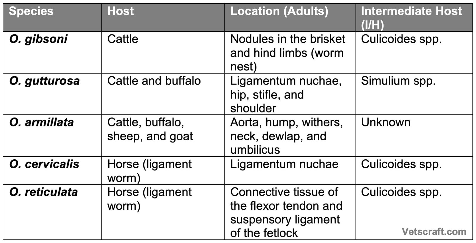
Taxonomical Classification
- Kingdom: Animalia
- Phylum: Nematoda
- Class: Chromadorea (Secernentea)
- Order: Spirurida
- Superfamily: Filarioidea
- Family: Onchocercidae
- Genus: Onchocerca
- Representative Species: Onchocerca volvulus, Onchocerca cervicalis, Onchocerca gibsoni, Onchocerca gutturosa, Onchocerca armillata, Onchocerca reticulata
- Common Name: Filarial worms (Onchocerca spp.)
Morphology
- Worms are elongate and filiform.
- Male worms measure 3–5 cm, while females measure 20–50 cm.
- Cuticle is transversely striated.
- Spicules are unequal and dissimilar.
- The tail end of the male is curved ventrally.
- Microfilariae are unsheathed.
Life Cycle
The adult worms live in nodules, and the fertilized females release microfilariae into the tissue lymphatic spaces, from where they are ingested by an insect vector, which acts as the intermediate host.
Development occurs in biting midges, and infective larvae are produced within 24–25 days. Cattle become infected when these biting flies carrying infective larvae feed on them.
Pathogenesis
- O. gibsoni: Causes “nodules” or “worm nests” in cattle, which contain coiled worms surrounded by a fibrous tissue capsule. The lesion gradually becomes thicker with age. Nodules may reach up to 5 cm in diameter and are ovoid or flat in shape. Later, they degenerate and undergo calcification or caseation. Affected animals usually do not show clinical signs, but infected carcasses are considered unfit for sale.
- O. gutturosa, O. cervicalis, O. lienalis: Adult worms are generally non-pathogenic. They live in tunnels and eventually degenerate. Such worms become surrounded by a granulomatous reaction. Heavy infections in horses may lead to lameness.
- O. armillata: Adult worms live in tunnels and nodules within the aorta. The aortic wall becomes thickened and contains numerous nodules filled with yellow caseous or slimy material containing coiled worms. In chronic cases, the nodules become calcified, and aneurysms may be seen.
Clinical Signs
Clinical signs include epileptiform episodes, blindness, and periodic ophthalmia.
Wahi/Kasen (Cattle) and Summer Mange/Allergic Dermatitis (Horses)
Wahi/Kasen (Cattle) and Summer Mange/Allergic Dermatitis (Horses) is a seasonal, sporadic dermatitis caused by microfilariae in the skin. It is characterized by papular or exudative dermatitis, alopecia, and severe pruritus.
The etiology is attributed to an immunological reaction against dead and dying microfilariae in the skin, along with an allergic reaction to insect bites. The microfilariae may also cause periodic ophthalmia, manifested by keratitis, conjunctivitis, keratouveitis, anterior uveitis, and blindness in horses.
In humans, O. volvulus causes ocular onchocerciasis (river blindness).
Diagnosis
- Adult parasites in the ligaments are detected only during postmortem examination.
- Nodules may be palpated.
- Skin biopsy for detection of microfilariae.
- Ophthalmic examination.
Treatment
Diethylcarbamazine: 5–8 mg/kg for 21 days. Systemic corticosteroids, metrifonate, trichlorfon, and ivermectin may also be used.
Control
Implement fly control measures and provide appropriate treatment to affected animals.

