
Guinea Worm (Dracunculus medinensis): Morphology, Life Cycle, Pathogenesis, Treatment & Control
Dracunculus medinensis, commonly known as the Guinea worm, is a tissue-dwelling parasitic nematode that causes dracunculiasis (Guinea worm disease). Humans are the principal definitive host, although dogs and some other mammals may also serve as definitive hosts in certain endemic regions. Infection occurs through the consumption of drinking water containing infected freshwater copepods (Cyclops spp.) that harbor the infective third-stage (L3) larvae.
This article provides a comprehensive overview of Dracunculus medinensis, including its taxonomy, morphology, transmission, life cycle, pathogenesis, clinical signs, treatment, and control measures. It is intended as a concise reference for veterinary and animal science students, veterinarians, and those preparing for competitive examinations.
Parasite Overview
- Common Name: Guinea worm, Medina worm, Serpent worm, and Dragon worm
- Predilection Site: Connective tissue and cutaneous swellings in humans, especially in the lower limbs
- Definitive Hosts: Humans (principal host); dogs and other mammals may serve as definitive hosts in some endemic regions
- Intermediate Hosts: Freshwater copepods (Cyclops spp.)
- Life Cycle: Indirect
- Infective Stage: Third-stage larvae (L3) within infected copepods (Cyclops spp.)
- Disease Caused: Dracunculiasis (Guinea worm disease)
Guinea worm (Dracunculus medinensis) is one of the oldest nematode parasites of humans. It was historically endemic in parts of India (including the Rajasthan desert) and Pakistan. It is also seen in dogs, cattle, and horses.
Taxonomical Classification
- Kingdom: Animalia
- Phylum: Nematoda
- Class: Chromadorea
- Order: Spirurida
- Superfamily: Dracunculoidea
- Family: Dracunculidae
- Genus: Dracunculus
- Species: Dracunculus medinensis
Morphology
Males are 10–30 mm long, whereas females are 300–400 cm long. In gravid females, the vulva becomes atrophied and is no longer functional.
Transmission
Infection occurs by drinking contaminated water containing Cyclops infected with the third-stage larvae of D. medinensis.
Life Cycle
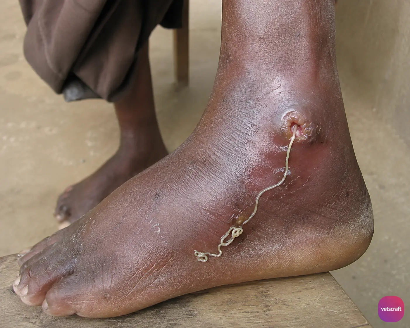
The adult worm lives in the subcutaneous (S/C) connective tissue, producing cutaneous swellings that develop into papules or blisters, mainly on the legs.
Later, the papule develops into an ulcer around the anterior extremity of the worm. When the infected person comes into contact with water, the uterus protrudes through the mouth of the worm and ruptures, releasing a large number of larvae (1–3 million) into the water.
The larvae are ingested by the intermediate host (I/H), Cyclops, in which they develop into the infective stage within 1–3 weeks. The free-living larvae survive for only 3 days in the absence of a host.
The definitive host (D/H) acquires the infection by ingesting infected I/H along with drinking water. The worm attains maturity in approximately one year.
Pathogenesis
- Female worms induce an allergic reaction followed by blister formation, usually on the lower limbs.
- Later, ulcer formation occurs.
- Burning sensation.
- Secondary bacterial infections may occur.
Clinical Signs
Urticaria, itching, and fever.
Treatment
- Thiabendazole: 50 mg/kg body weight for 2–3 days.
- Diethylcarbamazine, Niridazole: 25 mg/kg body weight for 10 days.
The best method of treatment is manual removal of the worm using a stick. During manual removal, the worm should not be broken. In this method, the worm is tied to a small stick and gradually rolled up over several days or weeks with great care.
Control
- Biological control by rearing fish.
- Use boiled drinking water.

