
Dictyocaulus filaria (Large Lungworm of Sheep and Goats): Complete Guide
Dictyocaulus filaria, commonly known as the large lungworm of sheep and goats, is an important nematode parasite of small ruminants. It primarily inhabits the trachea, bronchi, and bronchioles, where it causes dictyocaulosis, also referred to as verminous bronchitis or parasitic pneumonia. The disease is particularly significant in young animals and can lead to respiratory distress, reduced productivity, poor weight gain, and, in severe cases, death if left untreated.
This article provides a comprehensive overview of Dictyocaulus filaria, including its taxonomical classification, morphology, life cycle, pathogenesis, clinical signs, postmortem lesions, diagnosis, treatment, control, and immunoprophylaxis.
Parasite Overview
- Definitive Hosts: Sheep and goats
- Intermediate Host: None
- Life Cycle: Direct (monoxenous)
- Infective Stage: Third-stage larva (L3)
- Mode of Transmission: Ingestion of L3 larvae on contaminated pasture
- Predilection Site: Trachea, bronchi, and bronchioles of the lungs
- Disease Caused: Dictyocaulosis (verminous bronchitis or parasitic pneumonia)
Taxonomical Classification
- Kingdom: Animalia
- Phylum: Nematoda
- Class: Chromadorea
- Order: Rhabditida
- Suborder: Strongylina
- Superfamily: Trichostrongyloidea
- Family: Dictyocaulidae
- Genus: Dictyocaulus
- Species: Dictyocaulus filaria
- Common Name: Large lungworm of sheep and goats
Parasites of the family Dictyocaulidae inhabit the lungs and are therefore commonly referred to as lungworms.
Morphology
- The worms are milky white in color, and the intestine appears as a dark line.
- Female: 3–8 cm; Male: 5–10 cm in length.
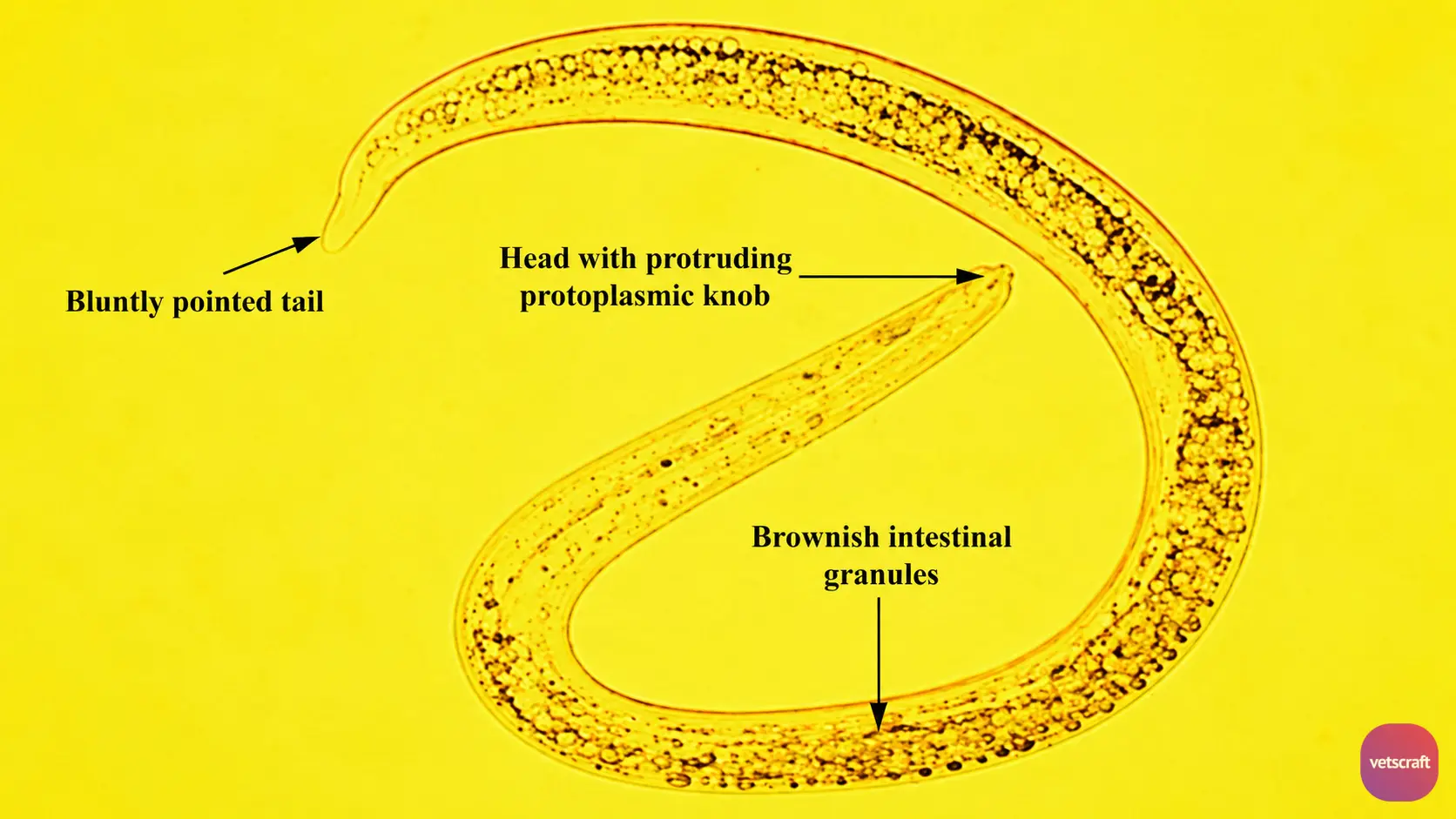
- The buccal capsule is shallow and surrounded by four lips. A knob is present on the head.
- In the male, the bursa is poorly developed. The mediolateral and posterolateral rays are fused except at their tips.
- The spicules are short, stout, reticulate, dark brown in color, equal in length, and boot- or sock-shaped.
- In the female, the vulva is situated just behind the midpoint of the body.
- The eggs contain fully developed larvae when laid.
- In the lungs, the worms become entangled, giving the appearance of “lumps of broken threads“.
Life Cycle
The eggs are coughed up and swallowed. The L1 larvae hatch from the eggs while passing through the intestine of the host and are voided in the feces. Occasionally, eggs may also be seen in sputum.
The L1 stage can be recognized by its cuticular knob at the anterior end and brownish food granules stored in the intestinal cells. Because of these stored food granules, the L1 larvae do not feed.
Within about 24 hours, the L1 molts to become L2. The L1 cuticle encloses the L2 until it molts to L3. Sometimes, the L3 may remain enclosed within two cuticles (L1 and L2).
The L2 molts to become L3 within 6 days.
The infective larvae climb onto grass blades during the early morning or under cool, humid conditions. Infection of the definitive host occurs through the ingestion of infective larvae along with herbage.
Following ingestion, the infective larvae penetrate the intestinal wall and enter the lymphatic vessels. They are carried by the lymph to the mesenteric lymph nodes, where the L3 larvae molt to become L4 within 4 days.
The L4 larvae reach the lungs via the lymphatic and blood vessels. In the lungs, they become lodged in the alveolar capillaries, then break through into the air passages and mature into adults within the bronchi in approximately four weeks.
Pathogenesis
The worms reside in the small bronchioles, causing parasitic catarrhal bronchitis. This condition is known as verminous pneumonia or verminous bronchitis. The inflammatory process extends into the surrounding peribronchial tissues, and the exudate spreads into the bronchioles and alveoli, resulting in atelectasis and pneumonia. Secondary bacterial infection may lead to severe pneumonia.
Clinical Signs
Young animals are primarily affected, although animals of all ages may be infected, and the disease is generally chronic. Coughing may be present but is not always observed.
Mucoid nasal discharge and dyspnea are common clinical signs. Increased respiratory rate and abnormal lung sounds may be detected on auscultation.
Postmortem Lesions
Areas of atelectasis of varying sizes are present. The affected bronchi contain adult worms and large amounts of mucus mixed with blood. The exudate appears opaque due to the presence of desquamated epithelial cells, leukocytes, and parasite eggs.
The bronchial mucosa and peribronchial tissues are inflamed and infiltrated with leukocytes. Localized, cone-shaped verminous pneumonia with consolidation of the lung parenchyma may be observed.
Compensatory emphysema and proliferation of the bronchial epithelium may occur. The lung lymph nodes are enlarged, and exudate accumulates within the bronchioles, resulting in collapse.
Diagnosis
Diagnosis is based on fecal examination for L1 larvae. Occasionally, eggs may also be detected in sputum or nasal discharge.
Treatment
- Diethylcarbamazine (DEC): 22 mg/kg/day for 3 days
- Methyridine: 200 mg/kg body weight, SC
- Tetramisole: 15 mg/kg body weight
- Levamisole: 7.5 mg/kg body weight, SC
- Fenbendazole: 5 mg/kg; Albendazole: 7.5 mg/kg
- Cyanacethydrazide: 15 mg/kg SC or 17.5 mg/kg orally
Control
- Remove animals from contaminated pastures.
- As larvae are commonly found in moist areas, animals should be allowed to graze on dry pastures.
- Provide a clean water supply.
Prevention (Immunoprophylaxis)
- Vaccination of animals.
- The vaccine consists of two doses of 1,000 X-ray-irradiated larvae each. Vaccination is performed at two months of age. The interval between the first and second doses is one month. Animals should be protected from natural infection until two weeks after the second dose.
- Reinfection after two weeks is necessary to maintain immunity.
- Dictol is a commercially available vaccine against lungworms that contains irradiated larvae.

