
Stephanofilaria in Animals: Classification, Pathogenesis, Diagnosis & Treatment
Stephanofilaria is a genus of filarial nematodes that causes cutaneous stephanofilariasis, a chronic parasitic skin disease affecting livestock, particularly cattle. The infection is characterized by dermatitis, ulcerative skin lesions, crust formation, and persistent irritation, leading to reduced animal welfare and economic losses. Depending on the species and geographic region, the disease is commonly referred to as humpsore in cattle.
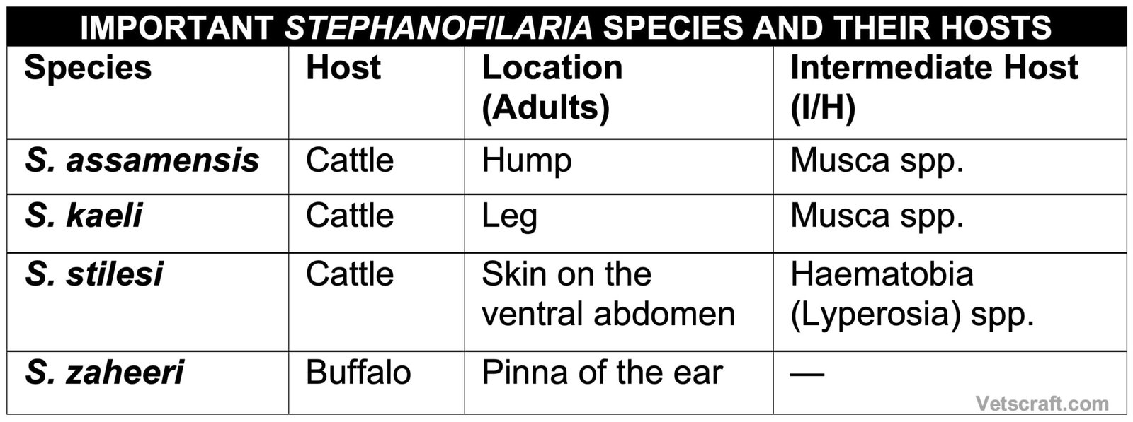
Several Stephanofilaria species have been reported worldwide, including S. assamensis, S. kaeli, S. stilesi, and S. zaheeri. These parasites inhabit the superficial layers of the skin and are transmitted by hematophagous flies that act as intermediate hosts. This article provides an overview of the taxonomy, pathogenesis, diagnosis, and treatment of Stephanofilaria infections in animals.
Stephanofilaria Species Overview
Taxonomical Classification
- Kingdom: Animalia
- Phylum: Nematoda
- Class: Chromadorea (Secernentea)
- Order: Spirurida
- Superfamily: Filarioidea
- Family: Filariidae
- Genus: Stephanofilaria
- Representative Species: Stephanofilaria assamensis, Stephanofilaria kaeli, Stephanofilaria stilesi, Stephanofilaria zaheeri
- Common Name: Stephanofilarial worm (causes cutaneous stephanofilariasis or humpsore in cattle)
Pathogenesis
Stephanofilaria causes humpsore (cutaneous stephanofilariasis) in cattle, which is a type of chronic dermatitis, also known as verminous dermatitis or Cascado.
Poor body condition and high rainfall are predisposing factors for this condition. Following infection, the lesions initially appear as a large number of small papules. Later, they coalesce to form a large lesion covered with a thick crust.
There are inflammatory changes, ulceration, acanthosis, hyperkeratosis, and alopecia. The lesions are rich in blood and lymph and can be readily expressed.
The lesion extends outward, while the center becomes hard and covered with a thick, dry crust. The lesion may reach up to 25 mm in diameter. Pruritus leads to constant rubbing against hard objects, and the condition is aggravated by self-inflicted wounds on the hump.
The worms live in the epithelial layers of the skin and cause inflammation of the rete malpighii, with proliferation and destruction of epithelial cells, hair follicles, and skin glands. The lesion contains both adult worms and microfilariae.
Secondary bacterial infection may occur. The lesions may partially resolve during the dry, cool season but reappear during the rainy season.
Diagnosis
- Diagnosis is based on microscopic examination of deep skin scrapings
- The scraping should be performed only after the crust has formed (Microfilariae can be detected)
Treatment
Apply trichlorfon ointment (6–10% in petroleum jelly or castor oil) for 7 days.

