
Habronema (Equine Stomach Worm): Morphology, Life Cycle, Pathogenesis, Diagnosis & Treatment
Habronema, commonly known as the equine stomach worm, is a spirurid nematode that primarily infects horses, donkeys, and other equids. The most important species include Habronema muscae, Habronema majus (H. microstoma), and Draschia megastoma. Adult worms inhabit the stomach, where they may cause chronic gastritis, gastric nodules, or fibrous tumors. Their life cycle involves flies, particularly Musca spp. and Stomoxys calcitrans, as intermediate hosts, making fly control an essential component of disease prevention.
In addition to gastric disease, infective larvae deposited on wounds or mucocutaneous junctions by flies can cause cutaneous habronemiasis (summer sores), a chronic granulomatous condition characterized by delayed wound healing and excessive granulation tissue. In this article, you’ll learn the taxonomy, morphology, life cycle, pathogenesis, clinical signs, diagnosis, treatment, and control of Habronema infections in horses.
Taxonomical Classification
- Kingdom: Animalia
- Phylum: Nematoda
- Class: Chromadorea (Secernentea)
- Order: Spirurida
- Superfamily: Habronematoidea
- Family: Habronematidae
- Genus: Habronema
- Important Species: Habronema muscae, Habronema majus (H. microstoma)
- Related Species: Draschia megastoma (formerly referred to as Habronema megastoma in older literature)
- Common Name: Equine Stomach Worm
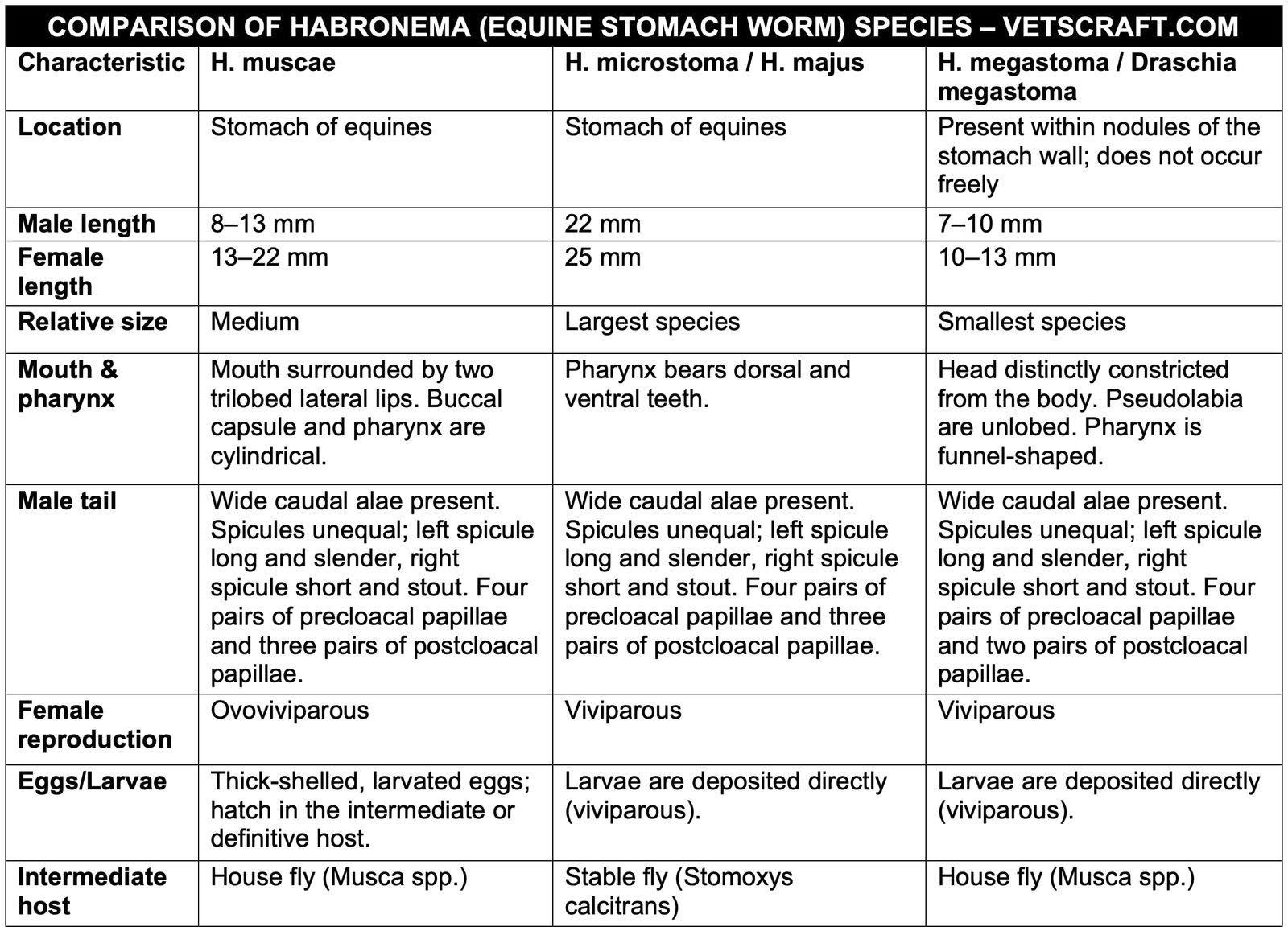
Morphology
Life Cycle of H. muscae and D. megastoma
Embryonated eggs or larvae are passed in the feces of the host (depending on the species). The eggs are ingested by the larvae of Musca spp. In the Musca larva, the L1 hatches and enters the Malpighian tubules, where it molts to become L2 by the time the Musca larva becomes a pupa.
In the pupa, the L2 molts to become L3. In the adult fly, the L3 migrates to the hemocoel (body cavity) and then to the proboscis. The infected adult fly deposits the larvae on the lips, nostrils, or existing wounds of the definitive host (D/H) while feeding. Following deposition, the L3 reaches the stomach of the horse and develops into the adult stage in approximately 2 months.
The larvae of the stable fly ingest L1. Development in the intermediate host (I/H) is similar to that of H. muscae. The normal feeding habit of Stomoxys calcitrans is biting and sucking.
Because the presence of infective larvae in the proboscis interferes with the fly’s feeding ability, the flies feed on moist surfaces such as the nostrils, lips, or existing wounds. The larvae deposited at these sites do not complete their development and remain localized within the wound, causing cutaneous habronemiasis.
Occasionally, flies may fall into water or feed, allowing horses to acquire infection by ingesting the larvae. The larvae mature into adults within approximately 2 months.
Pathogenesis of H. muscae and H. microstoma
These worms occur freely in the stomach and may penetrate the gastric mucosa, causing chronic catarrhal gastritis with excessive mucus production and ulceration of the gastric mucosa. H. microstoma may produce gastric ulcers.
Pathogenesis of D. megastoma
Adult worms produce large fibrous gastric tumors in the fundic region of the stomach wall of horses. The adult worms penetrate the gastric mucosa, causing nodule formation due to constant irritation. These tumors are frequently located in the fundic region and contain cavities filled with pus and caseous material in which the worms reside. The nodules usually have one or two openings.
In severe cases, multiple nodules may fuse to form a large tumor-like fibrous mass. This mass protrudes into the lumen of the stomach and causes mechanical interference with normal gastric function.
Clinical Signs
- Chronic gastritis.
- Tumors which located in the pyloric region may interfere with sphincter closure and result in digestive disturbances.
Summer Sore (Bursati, Swamp Cancer, Jack Sores, Granular Dermatitis, or Cutaneous Habronemiasis)
Summer Sore (Bursati, Swamp Cancer, Jack Sores, Granular Dermatitis, or Cutaneous Habronemiasis) is more common during summer because fly activity is greater than in winter. It is caused by the larvae of Habronema, particularly D. megastoma. Lesions are commonly observed on body parts that are prone to injury, such as the legs, withers, medial canthus of the eye, and sheath.
This condition occurs following the deposition of infective larvae on pre-existing wounds. The larvae cause irritation, resulting in delayed wound healing and the formation of reddish-brown material covered by granulation tissue.
The lesions gradually increase in size, do not respond to conventional treatment, and often heal spontaneously during winter.
In chronic cases, granulomatous lesions may develop. The larvae are also responsible for granular conjunctivitis. Lesions may occur on the medial canthus, the nictitating membrane, or the skin surrounding the eye, producing wart-like conjunctival lesions accompanied by epiphora and, occasionally, photophobia.
Larvae may also be found in the lungs, where they are associated with fibrotic nodules measuring 0.5–2 cm in diameter around the smaller bronchioles.
Diagnosis
Diagnosis is difficult and is generally based on the clinical appearance and behavior of the lesions.
Pruritus (itching), swelling, and excessive granulation tissue are characteristic findings. A biopsy is required for a definitive diagnosis.
Treatment
Surgical removal is recommended for cutaneous and ocular habronemiasis. Topical application of 10% chromic acid for 2–3 weeks may be used. Radiotherapy and cryosurgery are also effective for cutaneous and ocular habronemiasis.
Against adult worms:
- Fenbendazole: 15–60 mg/kg
- Trichlorfon: 25 mg/kg body weight
- Ivermectin: 200 µg/kg body weight
The animal should be fasted overnight. In the morning, administer 8–10 L of 2% NaHCO3 into the stomach using a stomach tube to loosen the gastric mucosa. After withdrawing the NaHCO3, administer carbon bisulfide at 5 mL/100 kg body weight, followed by a small quantity of water. This treatment is highly effective against adult worms.
Control
Properly dispose of manure and implement effective fly control measures.

