
Cooperia (Cattle Bankrupt Worm): Morphology, Life Cycle, Pathogenesis, Diagnosis & Treatment
Cooperia is a genus of gastrointestinal nematodes belonging to the family Cooperiidae. These parasites primarily infect the small intestine of domestic and wild ruminants, particularly cattle, sheep, and goats. Cooperia species are distributed worldwide and are among the most common parasitic nematodes affecting grazing livestock. Although infections are often subclinical, heavy worm burdens can reduce growth rates, impair feed efficiency, and cause significant economic losses in the livestock industry.
The life cycle of Cooperia is direct, with infection occurring through the ingestion of infective third-stage (L3) larvae from contaminated pasture. The pathogenic effects are generally mild compared with those caused by other trichostrongylid nematodes, but mixed infections with other gastrointestinal parasites can substantially increase disease severity.
The genus Cooperia comprises several species, of which the following are the most important in veterinary parasitology because of their prevalence and economic impact on domestic ruminants:
- Cooperia punctata – Cattle Cooperia Worm
- Cooperia pectinata – Cattle Cooperia Worm
- Cooperia oncophora – Intestinal Cooperia Worm (Cattle)
- Cooperia curticei – Sheep Cooperia Worm
- Cooperia surnabada – Cattle Cooperia Worm
Taxonomical Classification
- Kingdom: Animalia
- Phylum: Nematoda
- Class: Chromadorea (Secernentea)
- Order: Rhabditida (formerly Strongylida)
- Suborder: Strongylina
- Superfamily: Trichostrongyloidea
- Family: Cooperiidae
- Genus: Cooperia
- Common Name: Cattle Bankrupt Worm
Morphology
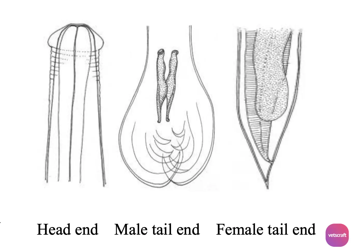
Members of the genus Cooperia are small, reddish nematodes. The anterior end is swollen due to the presence of a cephalic swelling, and the remainder of the body bears 14–16 longitudinal ridges that are transversely striated.
- The male bursa is large and has a small dorsal lobe.
- The spicules are short, stout, brown-pigmented, and possess a ridged, wing-like expansion in the middle.
- The gubernaculum is absent.
- The female has a vulvar flap and a tapered tail.
Life Cycle
Cooperia has a direct (monoxenous) life cycle and does not require an intermediate host. Adult worms inhabit the small intestine of ruminants, where they mate and produce eggs that are passed in the feces.
Under favorable environmental conditions, the eggs hatch into first-stage (L1) larvae, which feed on organic matter and molt into second-stage (L2) larvae. The L2 larvae then molt into infective third-stage (L3) larvae while retaining the cuticle of the previous stage as a protective sheath.
Ruminants become infected by ingesting sheathed L3 larvae while grazing on contaminated pasture. After ingestion, the larvae exsheath in the small intestine and penetrate the intestinal mucosa, where they develop into fourth-stage (L4) larvae.
Following further development, the immature adults emerge into the intestinal lumen, mature into adult worms, and begin producing eggs, thereby completing the life cycle. The prepatent period is generally 2–3 weeks, depending on the Cooperia species and environmental conditions.
Pathogenesis
The pathogenic effects of Cooperia result primarily from the development and feeding activity of the larval and adult stages in the small intestine.
During their development within the intestinal mucosa, the larvae cause mechanical damage to the intestinal epithelium, leading to villous atrophy, inflammation, and impaired absorptive function. Adult worms further contribute to mucosal irritation and disruption of normal intestinal physiology.
Light infections are usually subclinical, whereas heavy infections, particularly in young grazing cattle, can cause enteritis, reduced feed efficiency, poor weight gain, diarrhea, dehydration, and decreased productivity.
Mixed infections with other gastrointestinal nematodes, such as Ostertagia, Haemonchus, and Trichostrongylus, often exacerbate the severity of clinical disease and increase economic losses.
Diagnosis
Diagnosis is based on fecal examination and fecal culture. The infective third-stage (L3) larvae have a pointed tail enclosed within the retained sheath.
Treatment
- Benzimidazoles: Fenbendazole, Albendazole, Oxfendazole.
- Imidazothiazoles: Levamisole.
- Macrocyclic lactones: Ivermectin, Doramectin, Eprinomectin, and Moxidectin.
Selection of an anthelmintic should be based on the results of a fecal egg count reduction test (FECRT) or local anthelmintic resistance patterns, as Cooperia spp. commonly exhibit resistance to macrocyclic lactones.
Control
Control of Cooperia (Cattle Bankrupt Worm) relies on an integrated approach that combines strategic deworming, grazing management, refugia, FAMACHA scoring (where applicable), and other Integrated Parasite Management (IPM) practices. Since these control measures are common to most members of the Trichostrongylidae family, they are discussed in detail in our dedicated article: Trichostrongylidae Parasite Control: Refugia, FAMACHA & IPM.

