TABLE OF CONTENTS
Strongylus (Strongylus vulgaris, S. equinus & S. edentatus) in Horses: Complete Guide
Strongylus is a genus of large strongyles (large intestinal strongyles) that commonly parasitize horses and donkeys. The genus includes three important species: Strongylus vulgaris, Strongylus equinus, and Strongylus edentatus. Among these, S. vulgaris is considered the most pathogenic because its larval migration through the cranial mesenteric artery can cause severe vascular lesions, thromboembolism, and colic.
This article provides comprehensive veterinary notes on Strongylus, covering its taxonomic classification, egg morphology, life cycle, larval bionomics, pathogenesis, clinical signs, diagnosis, treatment, and control.
Parasite Overview
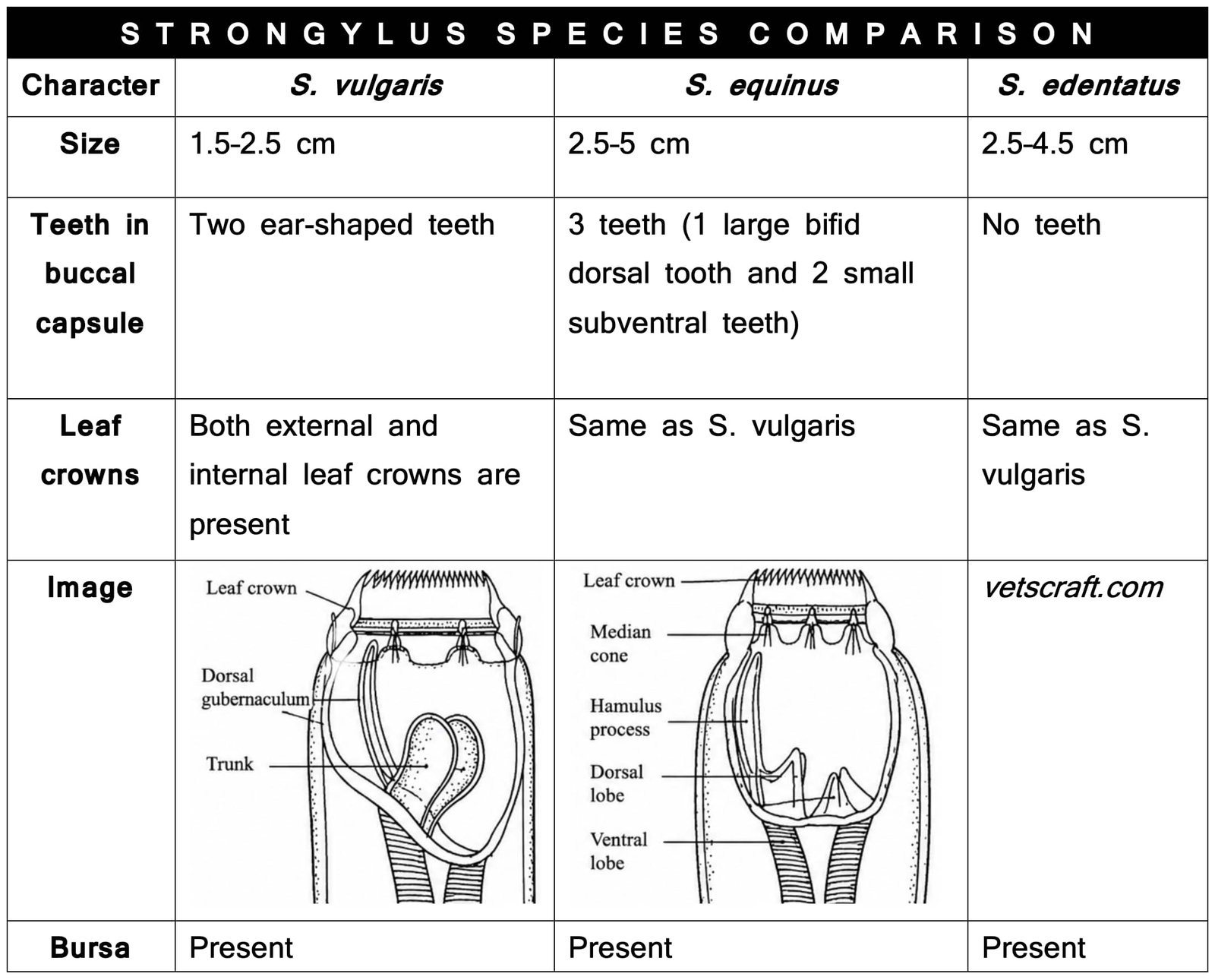
- Species: S. vulgaris, S. equinus, and S. edentatus
- Host: Horses and donkeys
- Location: Large intestine
- Infective Stage: L3 larvae
Taxonomical Classification of Strongylus vulgaris, Strongylus equinus, and Strongylus edentatus
- Kingdom: Animalia
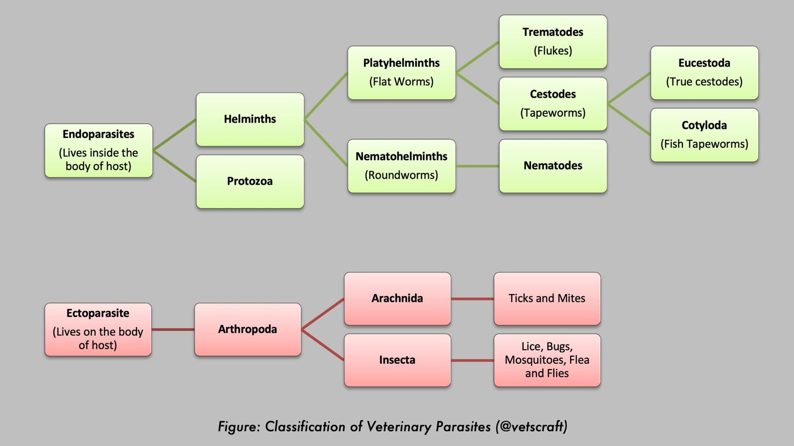
- Phylum: Nematoda
- Class: Chromadorea (Secernentea)
- Order: Strongylida
- Superfamily: Strongyloidea
- Family: Strongylidae
- Subfamily: Strongylinae
- Genus: Strongylus
- Species: Strongylus vulgaris, Strongylus equinus, and Strongylus edentatus
- Common Name: Large strongyles (large intestinal strongyles) of horses
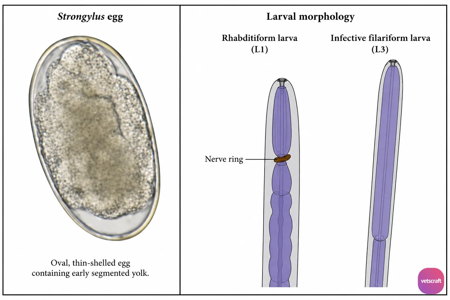
Egg
Eggs are oval in shape, thin-shelled, and contain an early segmented yolk.
Life Cycle
Eggs are passed in the feces of the host and contain an early segmented yolk. The development and hatching of eggs are controlled by various factors such as moisture, temperature, and O2. At 26°C, the first-stage larva is produced within 24 hours.
Bionomics of Strongyle Larva
- The L1 hatches from the egg. L1 has a rhabditiform esophagus. L1 mainly feeds on bacteria, grows rapidly, and moves very fast. It then enters a lethargic stage and molts to L2.
- L2 also feeds on bacteria. Soon after the lethargic stage, L2 molts to L3. L2 has a less developed rhabditiform esophagus.
- L3 has a filariform esophagus and is the infective stage. The cuticle of L2 is retained as a protective sheath around L3, which is very important for the survival of the infective stage.
- Since L3 is wrapped with the cuticle of L2, it does not feed. L3 survives on the stored food materials in the intestinal cells.
- The L3 larva is negatively geotropic and positively phototropic to mild sunlight but is repelled by strong sunlight. The response of L3 to heat and its migration are more active in warm weather than in cold weather.
- The L3 larva crawls up grass blades in the early morning, evening, and during cloudy weather. L3 can survive for up to 3 months.
- Horses become infected by ingesting L3 larvae along with herbage or vegetation.
- Exsheathment occurs in the small intestine of the horse.
- After exsheathment, L3 penetrates the intestinal wall, where it molts to become L4 within about 3 days of infection.
- Then L4 penetrates the intima of the submucosal arterioles and migrates toward the cranial mesenteric artery, where it produces thrombi and later aneurysms within about 14 days of infection.
- From 45 days after infection onward, L4 passes back to the submucosa of the cecum and colon via the arterial system. In the submucosa of the cecum and colon, L4 molts to L5. L5 then enters the intestinal lumen and reaches maturity in about 3 months.
Pathogenesis
The larval stages may be responsible for the more pathogenic effects.
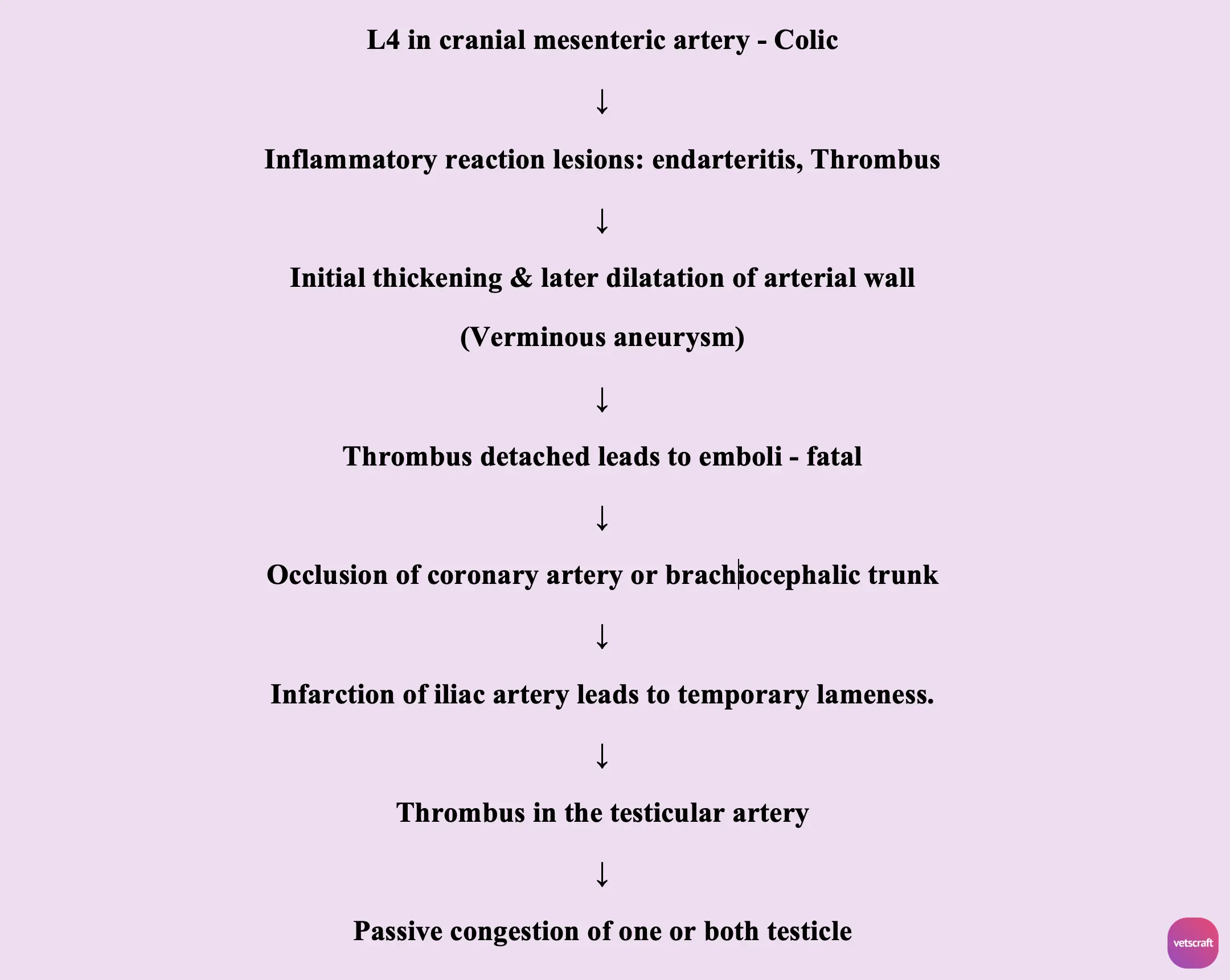
During the migration of L4 in the cranial mesenteric artery, it damages the endothelial cell lining and causes inflammatory lesions such as endarteritis and thrombus formation. Due to thrombus formation, initial thickening and later dilation of the arterial wall may occur. This condition is known as verminous aneurysm.
Sometimes the thrombus detaches and forms emboli. Occlusion of the coronary artery or brachiocephalic trunk may cause the death of the animal. Infarction of the iliac artery leads to temporary lameness. Thrombus formation in the testicular artery results in passive congestion of one or both testicles. A diarrheic syndrome may occur and is associated with ulceration of the cecum and colon due to thromboembolism caused by migrating larvae.
Due to Adult Worms
They attach to the mucosa, suck blood, and cause anemia (normocytic, normochromic anemia). They ingest plugs of mucosa, leading to the formation of small hemorrhagic ulcers. Later, these ulcers become confluent and produce ulcerated patches.
Clinical Signs
- Colic is due to pressure of the cranial mesenteric artery on the nerve plexuses caused by the migrating larvae and not by the presence of adult worms in the intestine.
- Rough hair coat, diminished appetite, diarrhea, edematous swelling of the abdomen and legs, emaciation, lameness, and anemia.
- Adult worms cause heavy blood loss due to their blood-sucking activity, resulting in anemia (normocytic, normochromic anemia).
Diagnosis
- Fecal examination for eggs.
- Detection of an aneurysm in the cranial mesenteric artery by rectal palpation.
- Postmortem examination reveals ascites, emaciation, and anemia. A large number of hemorrhagic ulcers are seen in the intestine, indicating the sites of worm attachment.
Treatment
- Fenbendazole: 7.5 mg/kg body weight (oral).
- Thiabendazole: 440 mg/kg body weight (oral).
- Ivermectin: 0.2 mg/kg body weight (S/C).
Control
- Deworming of horses.
- Proper disposal of manure.
- Use clean pastures.