TABLE OF CONTENTS
Taenia hydatigena: Morphology, Life Cycle, Pathogenesis & Diagnosis
Taenia hydatigena is a cyclophyllidean tapeworm of dogs and other wild carnivores, with domestic and wild ruminants serving as intermediate hosts. Its metacestode stage, Cysticercus tenuicollis, commonly develops on the omentum and peritoneal surfaces after migrating through the liver, where it can cause characteristic hemorrhagic tracts and traumatic hepatitis in young animals.
This article provides a concise overview of the taxonomy, morphology, life cycle, pathogenesis, and diagnosis of Taenia hydatigena for veterinary students and professionals.
Parasite Overview
- Host: Dogs and wild carnivores
- Location: Small intestine
- Intermediate Hosts: Domestic and wild ruminants (sheep, cattle, and occasionally pigs also act as intermediate hosts)
- Metacestode Stage: Cysticercus tenuicollis
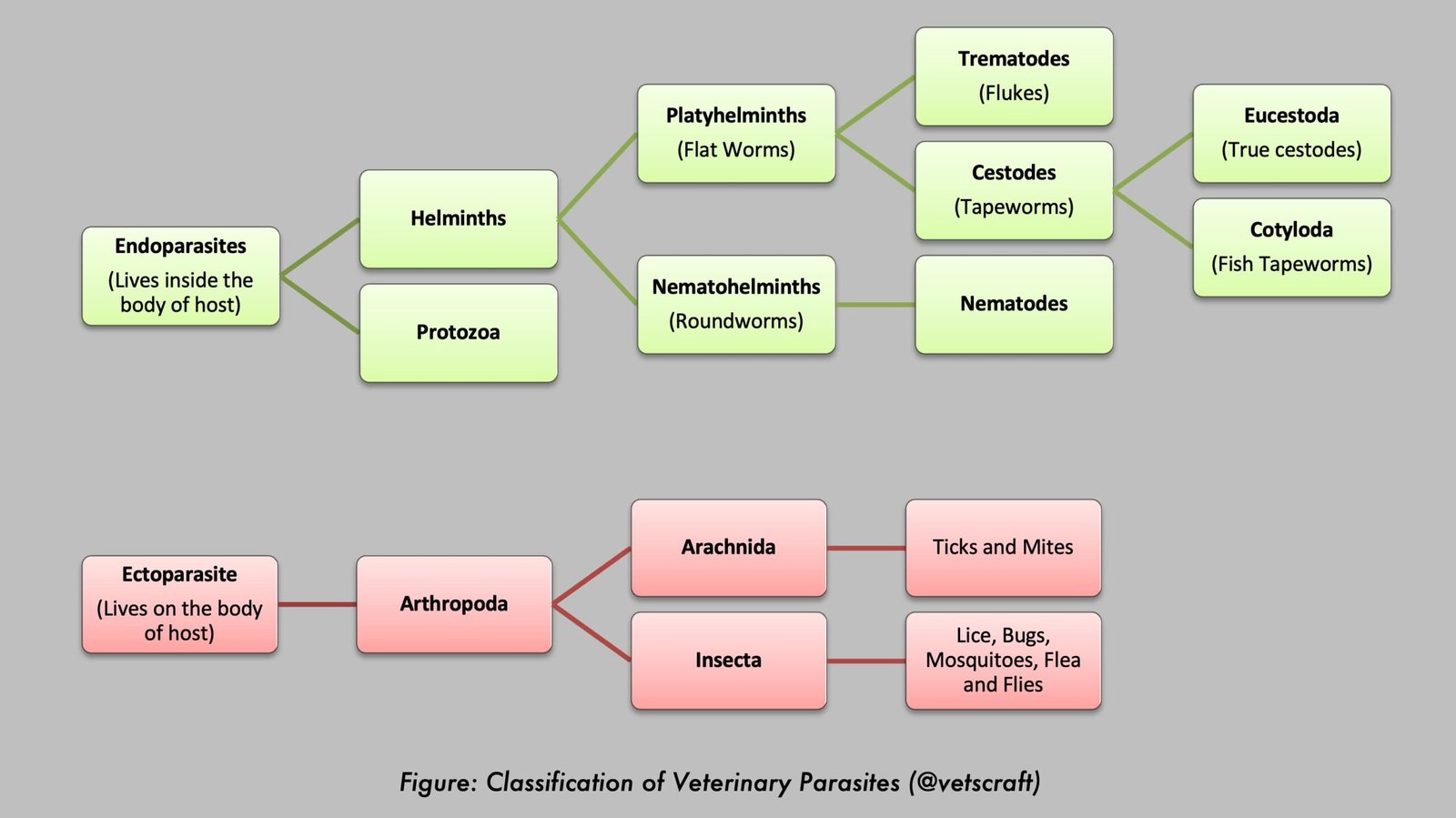
Taxonomical Classification
- Kingdom: Animalia
- Phylum: Platyhelminthes
- Class: Cestoda
- Subclass: Eucestoda
- Order: Cyclophyllidea
- Family: Taeniidae
- Genus: Taenia
- Species: Taenia hydatigena
- Common Name: Thin-necked bladder worm tapeworm
Morphology
- Rostellum armed with two rows of penknife-shaped hooks (1st row – 26 hooks; 2nd row – 46 hooks).
- Adults are up to 75–500 cm in length.
- Each segment contains a single set of genital organs. The genital pore is irregularly alternating.
- The ovary is situated at the posterior border of the segment.
- The uterus has a median stem.
- Testes are numerous.
- Gravid segments are longer than they are wide. In the gravid segment, the uterus has 6–10 lateral branches (an important characteristic for the identification of taeniid tapeworms).
Egg
- Eggs contain a hexacanth embryo.
- They have four layers composed of block-like structures, giving them a radially striated or “cartwheel” appearance.
Life Cycle
- Eggs hatch in the small intestine, releasing the hexacanth embryo.
- The hexacanth embryo penetrates the intestinal wall and reaches the liver via the circulation.
- In the liver, the hexacanth embryo breaks out of the portal vessels and migrates through the parenchyma for approximately one month. The developing cysticercus then migrates to the peritoneal cavity and reaches maturity in about 53 days. The mature cysticercus attaches to the omentum and mesenteric serosal surface of the intestinal wall. The metacestode stage is known as Cysticercus tenuicollis.
- Cysticercus tenuicollis: Approximately 6 cm in length, consisting of a single invaginated scolex attached to a fluid-filled bladder by a long neck. The definitive host acquires infection by ingesting meat or offal infected with Cysticercus tenuicollis.
- The prepatent period is approximately 51 days.
Pathogenesis
- The prevalence of infection is high in sheep, although the intensity of infection is generally low.
- Migration of Cysticercus tenuicollis through the liver causes hemorrhagic and fibrotic tracts. Heavy infections in lambs result in traumatic hepatitis, a condition known as hepatitis cysticercosa, which should be differentiated from acute fasciolosis. Cysticerci in the peritoneal cavity generally do not cause significant harmful effects.
Diagnosis
Diagnosis is made by postmortem examination of the intermediate host.