TABLE OF CONTENTS
Echinococcus granulosus in Dogs: Morphology, Life Cycle, Hydatid Cyst, Diagnosis & Treatment
Echinococcus granulosus is the smallest tapeworm of dogs and one of the most important zoonotic cestodes worldwide. Adult worms inhabit the small intestine of canids, while the larval stage develops as hydatid cysts in a wide range of intermediate hosts, including sheep, cattle, goats, pigs, horses, and humans.
The parasite causes cystic echinococcosis (hydatid disease), a significant public health and economic concern due to its chronic nature, potential organ damage, and zoonotic transmission. Understanding its morphology, life cycle, diagnosis, treatment, and control is essential for veterinarians, veterinary students, and public health professionals.
Parasite Overview
- Common Name: Smallest dog tapeworm (Important zoonotic tapeworm)
- Definitive Host: Dog
- Intermediate Hosts: All mammals, including humans
- Location: Small intestine
- Larval Stage: Hydatid cyst
Taxonomical Classification
- Kingdom: Animalia
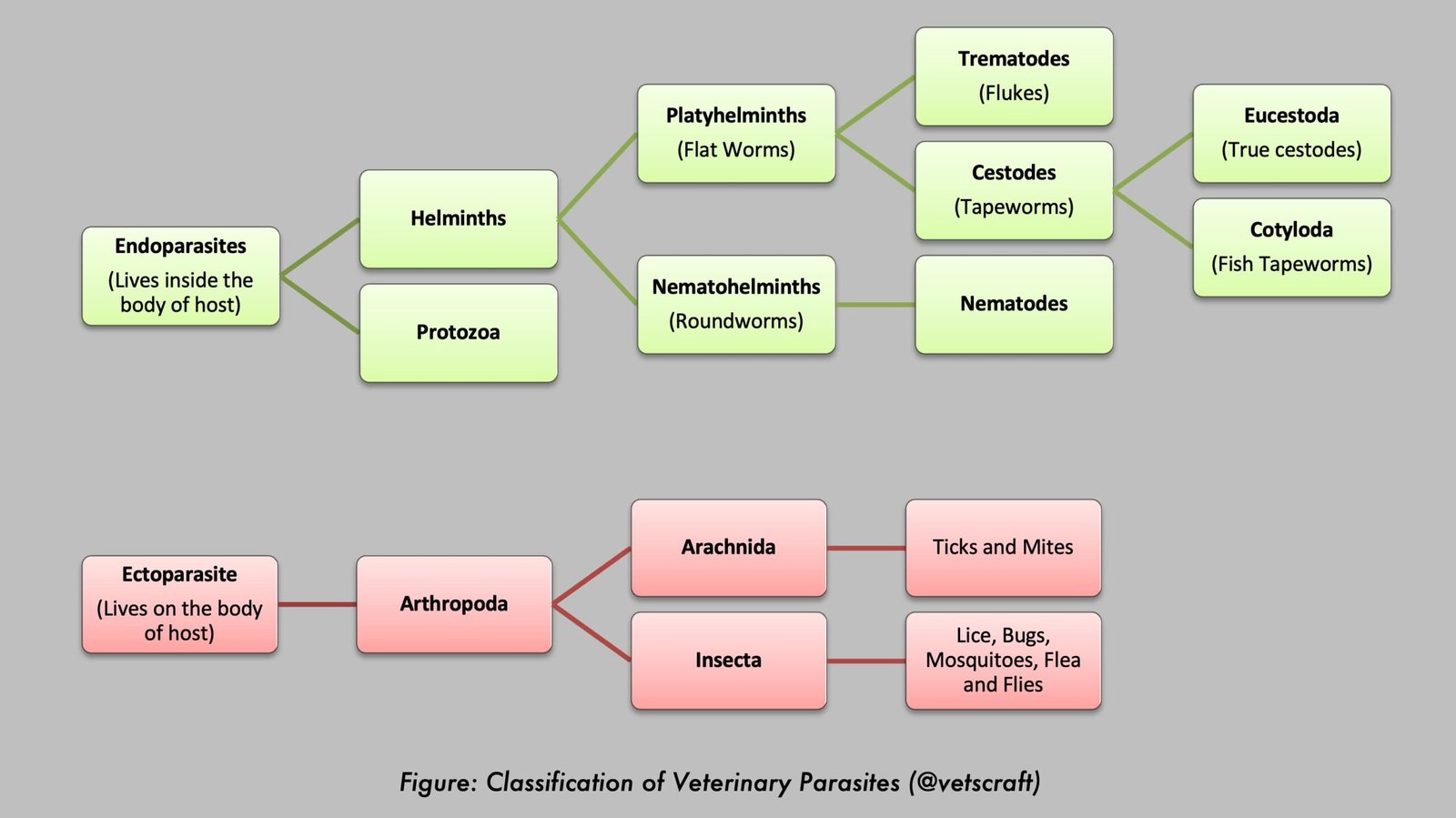
- Phylum: Platyhelminthes
- Class: Cestoda
- Subclass: Eucestoda
- Order: Cyclophyllidea
- Family: Taeniidae
- Genus: Echinococcus
- Species: Echinococcus granulosus
- Common Name: Hydatid tapeworm
Morphology
- The worms are 3 to 7 mm in length and have 3 to 4 segments.
- The rostellum has two rows of hooks.
- The penultimate segment is the mature segment, whereas the last segment is gravid.
- Each segment has a single set of reproductive organs.
- The genital pore alternates irregularly.
- The ovary is kidney-shaped. In the gravid segment, a number of lateral uterine branches may be present.
- Eggs are taeniid type.
Life Cycle
- Eggs are ingested by the intermediate host (sheep, cattle, goats, pigs, horses, and humans), where they hatch in the small intestine. The oncospheres penetrate the intestinal wall and reach the liver via the blood and lymphatic circulation.
- In the liver and lungs, the oncospheres develop into hydatid cysts.
- Cysts may also develop in other organs.
- Cysts develop slowly and require several months to reach maturity.
Hydatid Cyst
- Typically 5 to 10 cm in diameter, unilocular, and composed of two layers: an outer laminated membrane and an inner germinal membrane.
- Brood capsules develop from the germinal membrane approximately 5 months after infection.
- Each brood capsule contains numerous protoscolices.
- Sometimes, brood capsules detach and float freely in the hydatid fluid, forming what is known as “hydatid sand.”
- If the cyst ruptures, the brood capsules and protoscolices may produce external daughter cysts.
- Not all cysts produce brood capsules and protoscolices. Cysts lacking these structures are known as “sterile cysts.”
- Depending on the species, approximately 51% of hydatid cysts in sheep are sterile.
- The definitive host becomes infected by ingesting protoscolices present in infected meat or offal.
- In dogs, the protoscolices penetrate between the intestinal villi and reach maturity within approximately 4 to 7 days.
- Humans can acquire infection by ingesting eggs through contaminated food or by accidental entry of protoscolices through skin wounds during slaughter.
Pathogenesis
- Adult worms are generally non-pathogenic in dogs, whereas in humans and other domestic animals, severe disease is caused by the larval stage (hydatid cyst).
- Clinical signs depend on the location of the cyst. The function of the affected organ becomes impaired. If the cyst ruptures, it may result in anaphylactic shock.
Diagnosis in Dogs
- Fecal examination may reveal eggs; however, E. granulosus eggs cannot be differentiated from those of other taeniid species, such as T. multiceps and T. hydatigena.
- Definitive diagnosis is based on the demonstration of adult worms. To recover adult worms, the infected dog is treated with arecoline hydrobromide (1 to 2 mg/kg body weight). Following treatment, the dog passes its intestinal contents, including adult worms. The mucous portion of the final fecal material should be examined for the presence of adult worms.
Diagnosis in Humans (Hydatidosis)
- Casoni’s skin test
- Counterimmunoelectrophoresis
- ELISA
- AGPT
Treatment in Dogs
- Praziquantel: 5 mg/kg body weight orally or subcutaneously as a single dose. It is the treatment of choice and is highly effective against adult Echinococcus granulosus.
- Epsiprantel: 5.5 mg/kg body weight orally as a single dose. It is also effective against adult E. granulosus.
- Arecoline hydrobromide: Historically used at 1 to 2 mg/kg body weight for both diagnosis and treatment, but it is rarely recommended today because of its adverse effects and the availability of safer alternatives.
Treatment in Humans
- Surgical removal.
- PAIR technique (Puncture–Aspiration–Injection–Reaspiration)
- Aspiration of cyst fluid, marsupialization, and sterilization of the cyst.
- Injection of 2.5% to 10% formalin destroys the germinal membrane and protoscolices. However, this procedure is hazardous because spillage of cyst fluid may cause anaphylactic shock and dissemination of protoscolices to other parts of the body.
- Albendazole: 10 mg/kg body weight in two divided doses.
- Mebendazole: 400 to 600 mg/kg body weight, administered three times daily for 21 to 30 days.
Control of Tapeworm Infection in Dogs
- Control lice and fleas using 1% deltamethrin (Butox) or an appropriate flea collar.
- Maintain good kennel hygiene.
- Avoid feeding raw meat or offal to dogs.
- Perform routine deworming.
Control of Hydatid Cyst in Humans
- Maintain good personal hygiene.
- Public education.