
Ostertagia ostertagi (Brown Stomach Worm): Morphology, Life Cycle, Pathogenesis, Clinical Signs, Diagnosis & Treatment
Ostertagia ostertagi, commonly known as the brown stomach worm, is one of the most important gastrointestinal nematodes affecting cattle worldwide. It primarily inhabits the abomasum, where it damages the gastric glands, leading to impaired digestion, reduced productivity, diarrhea, weight loss, and significant economic losses in the cattle industry.
This article provides a comprehensive overview of Ostertagia ostertagi, including its taxonomy, morphology, life cycle, pathogenesis, clinical signs, diagnosis, and treatment. The content is designed for veterinary students, veterinarians, and competitive examination preparation.
Parasite Overview
- Common Name: Brown stomach worm
- Host: Sheep, goats, and cattle
- Predilection Site: Abomasum; rarely the small intestine
- Life Cycle: Direct life cycle (no intermediate host)
Taxonomical Classification
- Kingdom: Animalia
- Phylum: Nematoda
- Class: Chromadorea
- Order: Strongylida
- Superfamily: Trichostrongyloidea
- Family: Trichostrongylidae
- Subfamily: Ostertagiinae
- Genus: Ostertagia
- Species: Ostertagia ostertagi
- Common Name: Brown stomach worm of cattle
Morphology
- Worms are slender.
- The cuticle at the anterior end is slightly inflated and transversely striated.
- In females, the vulval flap is absent.
- Spicules are short, pigmented, with brown ends and 2/3 processes.
Life Cycle
- Adult worms inhabit the abomasum of ruminants and produce eggs that are passed in the feces.
- Under favorable environmental conditions, eggs hatch into first-stage larvae (L1), which develop into second-stage larvae (L2) and then into the infective third-stage larvae (L3) on pasture.
- Animals become infected by ingesting infective L3 larvae while grazing.
- After ingestion, L3 larvae exsheath in the rumen and migrate to the abomasum, where they penetrate the gastric glands.
- Within the gastric glands, larvae develop into fourth-stage larvae (L4) and then immature adults (L5).
- Immature adults emerge from the gastric glands into the lumen of the abomasum, where they mature into adult worms and begin egg production.
- Some L4 larvae may undergo hypobiosis (arrested development) within the gastric glands during unfavorable environmental conditions and resume development when conditions become favorable.
Pathogenesis
- Reduces the functional gastric gland mass and damages the gastric mucosa.
- Causes hyperplasia and thickening of the mucosa.
- Produces circular nodules measuring 2–3 mm in diameter, with a central orifice, due to extensive cell division and mucosal hyperplasia.
- In heavy infections, the mucosa becomes hyperplastic, giving a “Morocco leather” appearance. Oedema, necrosis, and sloughing of the abomasal mucosa occur, along with hypoalbuminemia due to increased permeability of the abomasal epithelium to pepsin and plasma proteins.
Clinical Signs
Abomasitis, oedema, necrosis, decreased albumin levels, anorexia, and profuse watery diarrhoea (bright green in color) due to the failure of the abomasum to denature chlorophyll. Morbidity is high.
The abomasal mucosa is thickened and oedematous, with oedema of the abomasum, decreased serum protein and albumin levels, and elevated plasma pepsinogen levels. Soiled hindquarters, anaemia, submandibular oedema, and loss of body weight are also observed.
Diagnosis
- Clinical signs, fecal examination, and fecal culture.
- Estimation of plasma pepsinogen levels.
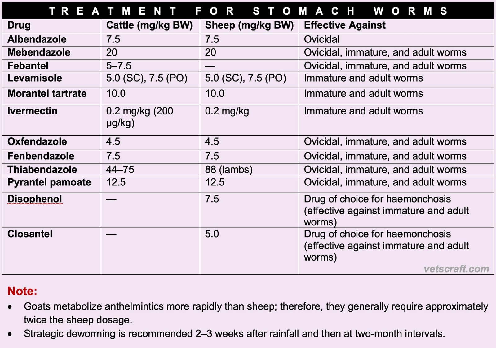
Treatment for Stomach Worms
Control
Control of Ostertagia ostertagi (Brown Stomach Worm) relies on an integrated approach that combines strategic deworming, grazing management, refugia, FAMACHA scoring (where applicable), and other Integrated Parasite Management (IPM) practices. Since these control measures are common to most members of the Trichostrongylidae family, they are discussed in detail in our dedicated article: Trichostrongylidae Parasite Control: Refugia, FAMACHA & IPM.

