TABLE OF CONTENTS
Oesophagostomum (Nodular Worm): Life Cycle, Pathogenesis, Clinical Signs & Treatment
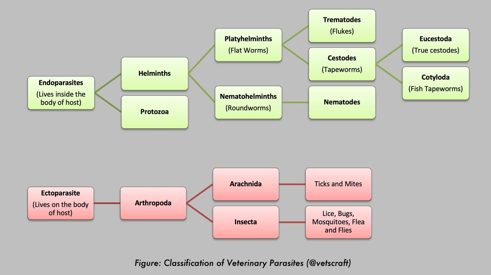
Oesophagostomum, commonly known as the nodular worm, is an important gastrointestinal nematode of domestic ruminants, particularly sheep, goats, and cattle. The parasite primarily inhabits the large intestine, where migrating larvae induce the formation of characteristic intestinal nodules, giving rise to conditions commonly referred to as pimply gut or knotty gut. Heavy infections can result in enteritis, persistent diarrhea, weight loss, poor growth, reduced wool production, and significant economic losses in livestock.
The two most important species are Oesophagostomum columbianum, which mainly infects sheep and goats, and Oesophagostomum radiatum, which primarily affects cattle. This article provides comprehensive veterinary notes on the taxonomy, life cycle, pathogenesis, clinical signs, diagnosis, treatment, control, and major differences between these two species.
Parasite Overview
- Oesophagostomum columbianum
- Host: Sheep, goats, camels
- Predilection Site: Large intestine (colon)
- Oesophagostomum radiatum
- Host: Cattle
- Predilection Site: Large intestine (colon)
Taxonomical Classification
- Kingdom: Animalia
- Phylum: Nematoda
- Class: Chromadorea
- Order: Strongylida
- Superfamily: Strongyloidea
- Family: Chabertiidae
- Subfamily: Oesophagostominae
- Genus: Oesophagostomum
- Species: Oesophagostomum columbianum, Oesophagostomum radiatum
- Common Names: Nodular worm of sheep and goats (O. columbianum), Nodular worm of cattle (O. radiatum)
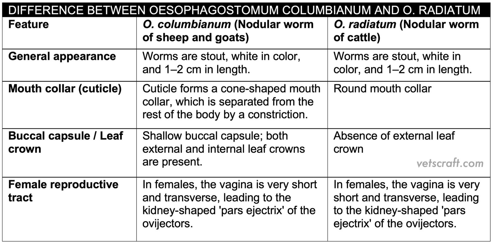
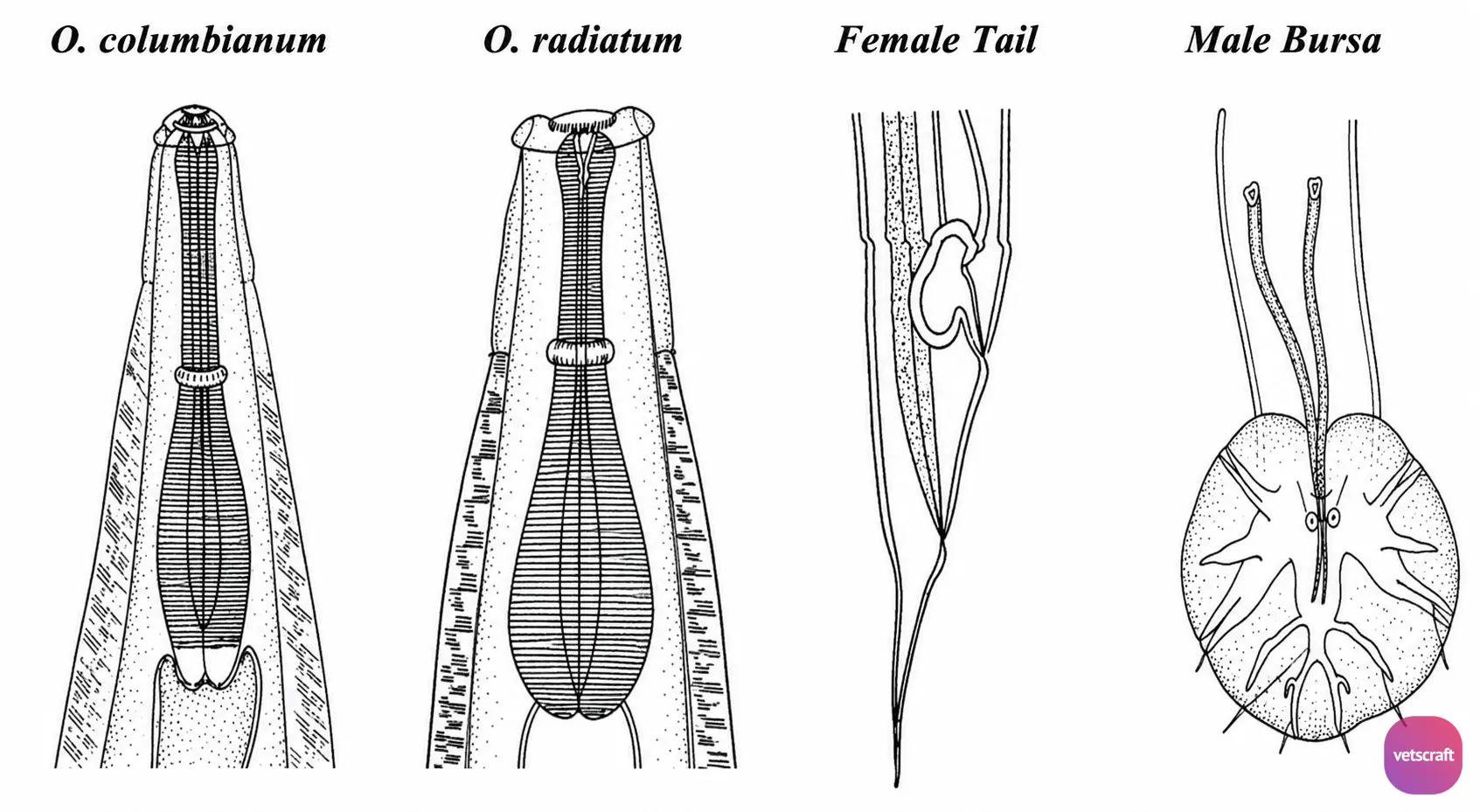
Difference Between O. columbianum and O. radiatum
Life Cycle
Eggs are of the strongyle type and are passed in the feces of the host. The development and bionomics of the larvae are similar to those of Strongyle spp.
L1 emerges from the eggs in about 20 hours, and L1 develops into L2 in about 3 days. The infective stage is reached in about 6–7 days. Infection of the final host occurs through the ingestion of infective larvae (L3) along with herbage. Exsheathment occurs in the small intestine.
L3 penetrates the intestinal wall from the pylorus to the rectum, coils within the muscularis mucosae, and forms a nodule (cyst), where L3 molts into L4 in about 4 days. The L4 then enters the intestinal lumen and passes to the colon, where it molts into L5 and reaches maturity in approximately 41 days.
Oesophagostomum columbianum
Oesophagostomum columbianum is the principal nodular worm of sheep and goats and is one of the most pathogenic intestinal nematodes of small ruminants. The larvae migrate through the intestinal wall, producing characteristic nodules that may lead to severe enteritis, diarrhea, weight loss, and significant economic losses in affected flocks.
Pathogenesis of O. columbianum
It is a serious pathogen of sheep. Approximately 200–300 adult worms can cause severe infection in young sheep. In the intestine, they produce a condition known as pimply gut or knotty gut. Lambs and older sheep exposed to this parasite for the first time have no prior immunity; therefore, the larvae induce little to no reaction during migration through the intestinal mucosa. Eventually, large numbers of adult worms are present in the colon, but no nodules are formed in the intestinal wall.
In contrast, in lambs and sheep with previous exposure to this parasite (i.e., immunologically sensitized sheep), the larvae produce a localized reaction during migration through the mucosa. During this localized reaction, leukocytes, especially eosinophils, and foreign-body giant cells accumulate around each larva and become encapsulated by fibroblasts. The larvae remain within the nodules for about three months.
When the contents of the nodules undergo caseation and calcification, the larvae either die or leave the nodules. After leaving the nodules, the larvae wander through the muscle fibers. However, the majority of the larvae do not return to the intestinal lumen. In such cases, large numbers of nodules and very few adult worms are observed. Nodules may rupture into the peritoneal cavity, causing peritonitis and multiple adhesions.
Nodules in the small or large intestine interfere with absorption, bowel movement, and digestion. They cause thickening of the intestinal wall, congestion, excessive mucus production, and hypoalbuminemia. As a result, body growth and wool production are reduced.
Economic Importance
Because the nodular intestine is unfit for human consumption, it must be condemned during slaughter. It is also unsuitable for catgut production.
Clinical Signs
- Marked and persistent diarrhea. The feces are dark green in color and mixed with mucus, sometimes containing blood. The diarrheic syndrome occurs about 6 days after infection and coincides with the time when the larvae leave the nodules.
- In chronic cases, diarrhea may occur initially, followed by constipation, along with extreme emaciation, cachexia, general weakness, dry skin, poor-quality wool, and muscle atrophy, eventually leading to complete prostration for 1–3 days and death.
Postmortem Lesions
Marked emaciation with a complete absence of body fat. In the initial stage of infection, large numbers of worms are present, and the mucosa is thickened, reddened, and covered with mucus. In repeated infections, the intestinal walls are studded with nodules, some of which develop into abscesses containing green to yellowish pus or caseous material.
Diagnosis
Fecal examination for eggs.
Treatment
- Benzimidazole Compounds:
- Fenbendazole: 7.5 mg/kg b.wt.
- Thiabendazole: 44 mg/kg b.wt.
- Albendazole: 5–10 mg/kg b.wt.
- Piperazine Compounds: 5–15 mg/50 kg b.wt.
- Levamisole: 7.5 mg/kg b.wt.
- Supportive therapy may be necessary in severe cases of diarrhea.
Control
Maintain clean pastures and perform periodic deworming.
Oesophagostomum radiatum
Oesophagostomum radiatum is the nodular worm of cattle and primarily inhabits the large intestine. Although adult cattle usually tolerate light infections, heavy worm burdens, especially in young animals, can cause enteritis, diarrhea, poor growth, and reduced productivity.
Pathogenesis of O. radiatum in Cattle
O. radiatum is more pathogenic in cattle when present in large numbers. In acute cases, inflammation of the small and large intestines and black, foul-smelling diarrhea are observed. Chronic cases are more severe in young animals, whereas adults usually recover.
In the intestine, it produces a condition known as pimply gut or knotty gut, which is associated with intermittent diarrhea that later progresses to purging, resulting in emaciation, prostration, and death in young animals. Normocytic anemia and hypoproteinemia occur due to protein-losing enteropathy.
Diagnosis, Treatment, and Control
The diagnosis, treatment, and control of O. radiatum infection are similar to those of O. columbianum infection.