
Nematodirus spathiger (Thread-Necked Strongyle Worm)
Nematodirus spathiger is a gastrointestinal nematode of the family Molineidae that primarily infects the small intestine of domestic and wild ruminants, particularly sheep and goats. It is commonly referred to as the thread-necked intestinal worm or thread-necked strongyle worm because of its characteristically slender anterior end.
This parasite is of veterinary importance due to its ability to cause enteritis, villous atrophy, diarrhea, dehydration, reduced nutrient absorption, poor weight gain, and production losses in infected animals, especially young lambs and kids. Understanding its morphology, life cycle, pathogenesis, clinical signs, and diagnosis is essential for effective prevention and control.
Taxonomical Classification
- Kingdom: Animalia
- Phylum: Nematoda
- Class: Chromadorea
- Order: Strongylida
- Superfamily: Trichostrongyloidea
- Family: Molineidae
- Subfamily: Nematodirinae
- Genus: Nematodirus
- Species: Nematodirus spathiger
- Common Name: Thread-necked intestinal worm
Morphology
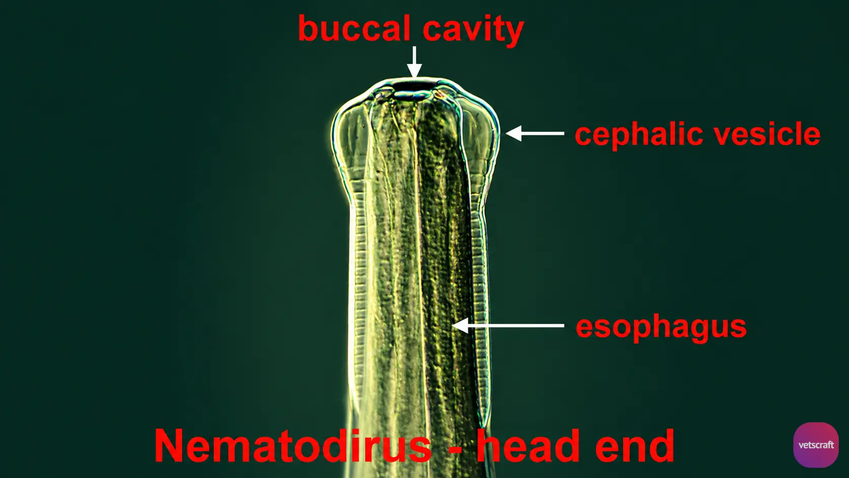
- Members of this genus are long with a slender anterior portion.
- The inflated cuticle at the anterior end has 14–18 longitudinal ridges.
- The male bursa has well-developed lateral lobes, whereas the dorsal lobe is indistinct.
- Spicules are long and slender, with fused tips.
- The tail of the female is short and bears a terminal appendage.
- The eggs are much larger than those of other strongyle species.
Life Cycle
Eggs are passed in the feces of the host. After two to three months, the infective L3 develops within the egg. The final host becomes infected by ingesting infective larvae along with vegetation or by ingesting eggs containing L3 larvae.
Following ingestion, exsheathment occurs in the intestine. The infective larvae penetrate the intestinal mucosa, where L3 molts to L4 in about 5 days, and L4 molts to L5 in about 5 days. Finally, L5 returns to the intestinal lumen and reaches maturity.
Pathogenesis
In the small intestine, extensive destruction of the ileal mucosa occurs. The parasite forms tunnels through the mucosa, with the posterior part protruding outside. By the 16th day, the villous surface is reduced, and villous atrophy is evident by the 20th day. The intestinal mucosa returns to normal by the 32nd day. The absorptive capacity of the intestine for nutrients and fluids is reduced.
Clinical Signs
Diarrhea, emaciation, dehydration, and inflammation of the intestinal mucosa are common. Animals become thirsty and continue to graze while congregating around drinking areas.
Diagnosis
Diagnosis is based on fecal examination (large-sized eggs) and fecal culture.

