
Ancylostoma (Hookworm): Morphology, Life Cycle, Pathogenesis, Clinical Signs, Diagnosis & Treatment
Ancylostoma, commonly known as hookworm, is a genus of blood-feeding gastrointestinal nematodes that infects dogs, cats, wild carnivores, and occasionally humans. These parasites are of major veterinary importance because they cause severe anemia, enteritis, poor growth, and even death in young animals. Infection occurs through ingestion of infective larvae, skin penetration, transmammary transmission, or paratenic hosts, depending on the species. In addition to their veterinary significance, some Ancylostoma species are zoonotic and can cause cutaneous larva migrans (CLM) in humans. This article covers the taxonomy, morphology, life cycle, pathogenesis, clinical signs, diagnosis, treatment, and control of Ancylostoma (hookworm) in veterinary parasitology.
Several species of Ancylostoma have been identified in domestic animals and humans. Among these, a few species are of major veterinary importance because they infect dogs and cats, while others are primarily associated with human infections. The following are the most important species of Ancylostoma recognized in veterinary parasitology:
- Ancylostoma caninum: Hookworm of dogs
- Ancylostoma braziliense: Hookworm of dogs and cats
- Ancylostoma tubaeforme: Hookworm of cats
- Ancylostoma ceylanicum: Hookworm of dogs and cats (zoonotic)
- Ancylostoma duodenale: Human hookworm (occasionally included for comparison)
Taxonomical Classification
- Kingdom: Animalia
- Phylum: Nematoda
- Class: Chromadorea (Secernentea)
- Order: Rhabditida (formerly Strongylida)
- Suborder: Strongylina
- Superfamily: Ancylostomatoidea
- Family: Ancylostomatidae
- Subfamily: Ancylostominae
- Genus: Ancylostoma
- Common Name: Hookworm
Morphology
The anterior end of the worm is bent dorsally, giving the parasite its characteristic hook-like appearance; hence the name “hookworm.” They possess a well-developed, subglobular buccal capsule. Leaf crowns are absent. Depending on the species, the buccal capsule contains either teeth or cutting plates. They are voracious bloodsuckers.
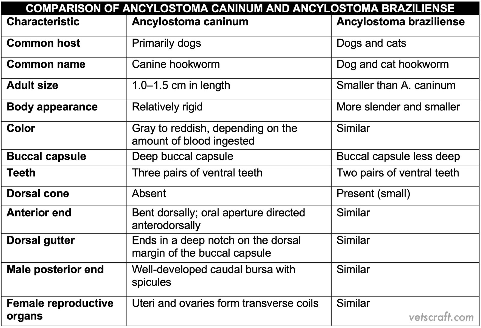
Comparison of A. caninum and A. braziliense
Life Cycle
Females lay approximately 16,000 eggs per day. The bionomics and larval development are similar to those of Strongyle spp. Desiccation is highly lethal to hookworm larvae; therefore, slightly sandy, moist soil provides the most favorable environment for their survival.
The first-stage larva (L1) emerges from the egg in approximately 3 days and molts to become L2 within 6 days. Under optimal conditions (23–30°C), the infective L3 stage is reached within 1 week. Infection of the definitive host occurs either by oral ingestion of L3 larvae or by skin penetration of L3 larvae.
In dogs and cats, oral ingestion is the most common route of infection because of their habit of licking grass. Skin penetration is more common in humans and cattle.
In A. caninum, infection occurs by five routes:
- Oral ingestion
- Skin penetration
- Prenatal infection
- Transmammary infection
- Paratenic host transmission
1. Oral Ingestion
Following oral ingestion, the infective larvae either develop directly into adults or penetrate the oral mucosa and migrate via the tracheal route. Ultimately, they reach the intestine, where they develop into adults.
2. Skin Penetration
Following skin penetration, the infective larvae reach the lungs via the bloodstream and then migrate through the tracheal route. They subsequently enter the intestine, where they develop into adults.
3. Prenatal Infection
In older bitches, following oral ingestion or skin penetration, a few larvae develop directly into adults, whereas the majority undergo somatic migration and remain dormant within the tissues until pregnancy. During pregnancy, these dormant larvae are reactivated by hormonal influences and migrate to the fetus via the placental circulation.
The worms do not mature until after the pups are born. They mature within 30 days after birth, and eggs can then be detected in the feces. Prenatal infection is common in puppies (similar to T. canis).
4. Transmammary Infection
The larvae are transmitted to puppies through colostrum, where they develop directly into adults without undergoing tissue migration.
5. Paratenic Host
Paratenic hosts, such as rodents, may also be involved. Infection occurs through the ingestion of infected rodents.
Pathogenesis
Anemia is the principal clinical sign of A. caninum infection, and it is considered one of the most pathogenic parasites of dogs. Heavy infections commonly occur in puppies less than 1 year of age. Smaller breeds are more severely affected than larger breeds.
The worms attach to the intestinal mucosa using their well-developed buccal capsule and suck blood. They frequently change their site of attachment, resulting in numerous necrotic foci. Because the worms secrete anticoagulants, blood continues to ooze from the previous attachment sites.
In heavy infections, puppies rapidly become anemic because each worm consumes approximately 0.01–0.09 mL of blood per day, depending on the severity of infection. The development of anemia coincides with the emergence of the L5 stage, as it possesses the adult buccal capsule. Initially, the anemia is normocytic and normochromic; however, as the animal becomes iron deficient, a microcytic hypochromic anemia develops.
In dogs, skin penetration by L3 larvae causes moist eczema, ulceration, and swelling of the subcutaneous tissues. Larval migration also results in decreased RBC and hemoglobin levels, pulmonary hemorrhage, and hemorrhagic pneumonitis. In chronic infections, reduced appetite, a poor hair coat, and poor growth are commonly observed. Anemia is generally not observed in A. braziliense infection because the associated blood loss is insignificant.
Clinical Signs
Anemia depends on the age of the host, nutritional status, and iron reserves. Severe anemia may lead to coma and death within 3 weeks after birth. Hydremia, edema, weakness, and emaciation may also occur. Eosinophilia, a dry and harsh hair coat, and stunted growth are common findings.
Skin irritation and dermatitis occur due to larval penetration. The mucous membranes become pale. Affected animals may develop diarrhea containing blood and mucus and pass dark, tarry feces (melena).
Cutaneous Larva Migrans (CLM), also known as Creeping Eruption, is a condition that occurs in unnatural hosts, such as humans (especially children), following skin penetration by the larvae of A. braziliense, A. caninum, A. duodenale, Uncinaria stenocephala, Necator americanus, Bunostomum phlebotomum, Strongyloides spp., and Gnathostoma spinigerum.
Immunity
Dogs gradually develop immunity to infection. Graded doses of larvae administered orally or subcutaneously can induce protective immunity. Egg counts increase following the initial infection, reaching a peak approximately 2 months post-infection. Thereafter, egg counts decline, and large numbers of adult worms are expelled in the feces. This phenomenon is referred to as a “crisis” and is comparable to the self-cure phenomenon observed in H. contortus infection.
Diagnosis
- Clinical signs and fecal examination in puppies.
- Severe anemia in puppies.
Treatment
- Bephenium bromide or iodide: 20 mg/kg body weight
- Mebendazole: 40 mg/kg body weight
- Dichlorvos: 12–15 mg/kg body weight
- Tetramisole:
- 10 mg/kg body weight (SC injection)
- 20 mg/kg body weight (oral administration)
- Fenbendazole: 7.5 mg/kg body weight
- Thiabendazole: 20 mg/kg body weight
- Disophenol: 7.5 mg/kg body weight (SC injection)
Control
- Treat kennel floors with common salt or a sodium borate solution.
- Implement periodic deworming of puppies.
- Maintain proper kennel hygiene.
- Ensure proper disposal of feces.

