
Setaria in Animals: Morphology, Life Cycle, Pathogenesis & Treatment
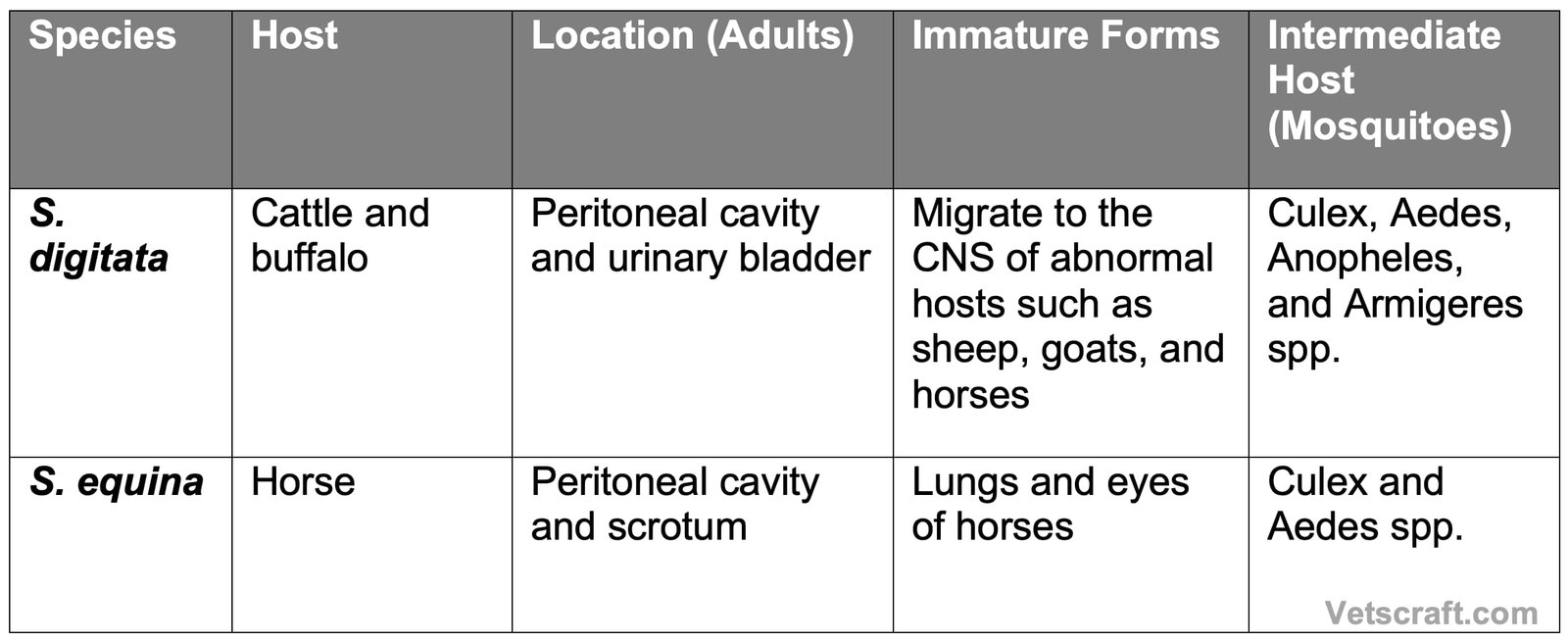
Setaria species are filarial nematodes that commonly inhabit the peritoneal cavity of domestic and wild ungulates. Adult worms are generally non-pathogenic in their natural hosts; however, the migrating larval stages may cause severe neurological disease, known as enzootic cerebrospinal nematodiasis (Kumri or lumbar paralysis), in abnormal hosts such as sheep, goats, and horses. These parasites are transmitted by mosquitoes, which serve as intermediate hosts and play an essential role in their life cycle.
Setaria Species Overview
Taxonomical Classification of Setaria
- Kingdom: Animalia
- Phylum: Nematoda
- Class: Chromadorea
- Order: Spirurida
- Superfamily: Filarioidea
- Family: Onchocercidae
- Genus: Setaria
- Important Species: Setaria cervi, Setaria digitata, Setaria equina, Setaria labiatopapillosa, Setaria marshalli
- Common Name: Abdominal (Peritoneal) filarial worms
Morphology
- Worms are long and milky white in color.
- The mouth is surrounded by a chitinous ring called the “peribuccal ring.” It has lateral, ventral, and dorsal prominences.
- The body of the worm tapers toward the tail end, and the posterior end is spirally coiled.
- Spicules are unequal and dissimilar.
- The tail end of both sexes has small lateral appendages.
- The female tail end has a small knob-like conical projection and is armed with a small spine.
- The microfilaria is sheathed.
Life Cycle
The exact life cycle of setaria is unknown. However, development occurs in the thoracic cavity of mosquitoes, and infective larvae are produced within 12–16 days.
Pathogenesis
The adult parasite is generally non-pathogenic, but in some cases it causes fibrinous peritonitis. S. digitata causes eosinophilic granulomatous lesions in the urinary bladder. In rare cases, adult S. digitata may undergo erratic migration into the eyes of horses, causing blindness.
Enzootic Cerebrospinal Nematodiasis (Kumri or Lumbar Paralysis)
Enzootic cerebrospinal nematodiasis (Kumri or lumbar paralysis) is a condition caused by the erratic migration of the immature stages of S. digitata in unnatural hosts such as horses, sheep, and goats.
The condition is more prevalent during summer because mosquito activity is higher. Affected animals develop acute focal encephalomyelomalacia.
The migrating worms produce tracks that cause acute malacia with disintegration of all tissues at the center of the lesion and degeneration of nerve tracts.
Clinical Signs
- Clinical signs depend on the site of the lesion.
- Muscular weakness, ataxia, paralysis of the hind limbs, and sometimes blindness may occur.
Treatment
DEC: 40 mg/kg body weight for 1–3 days.

