TABLE OF CONTENTS
Conjunctivitis in animals
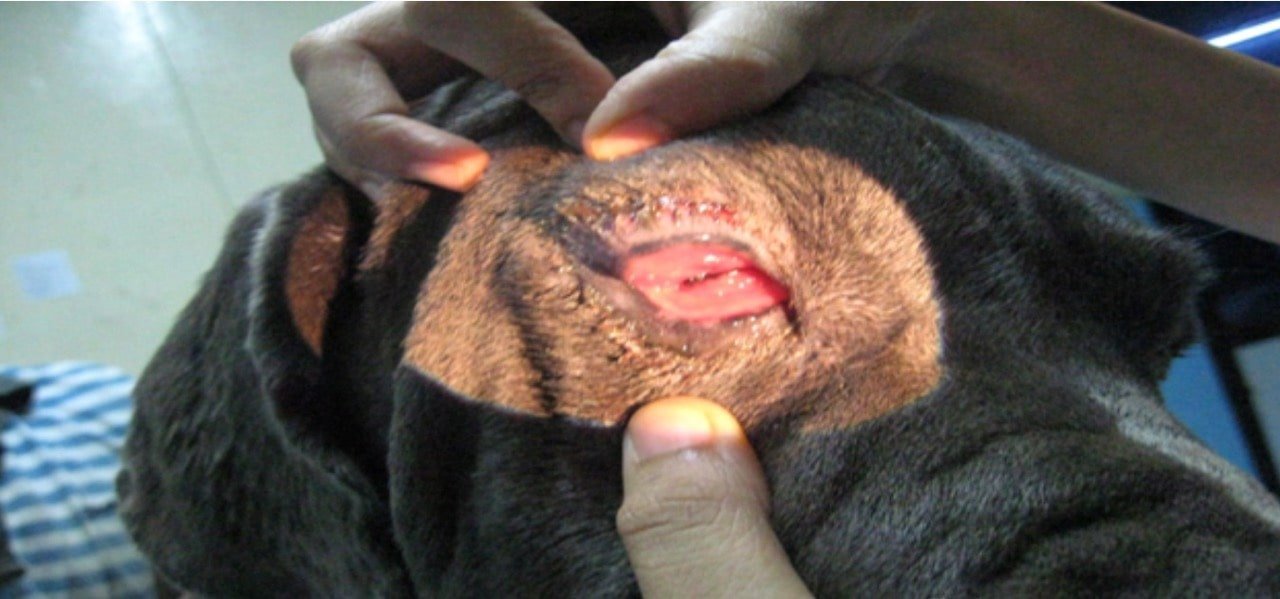
Conjunctivitis is inflammation of the conjunctiva is one of the most common eye diseases in animals.
Conjunctiva has two parts called palpebral conjunctiva and the bulbar conjunctiva.
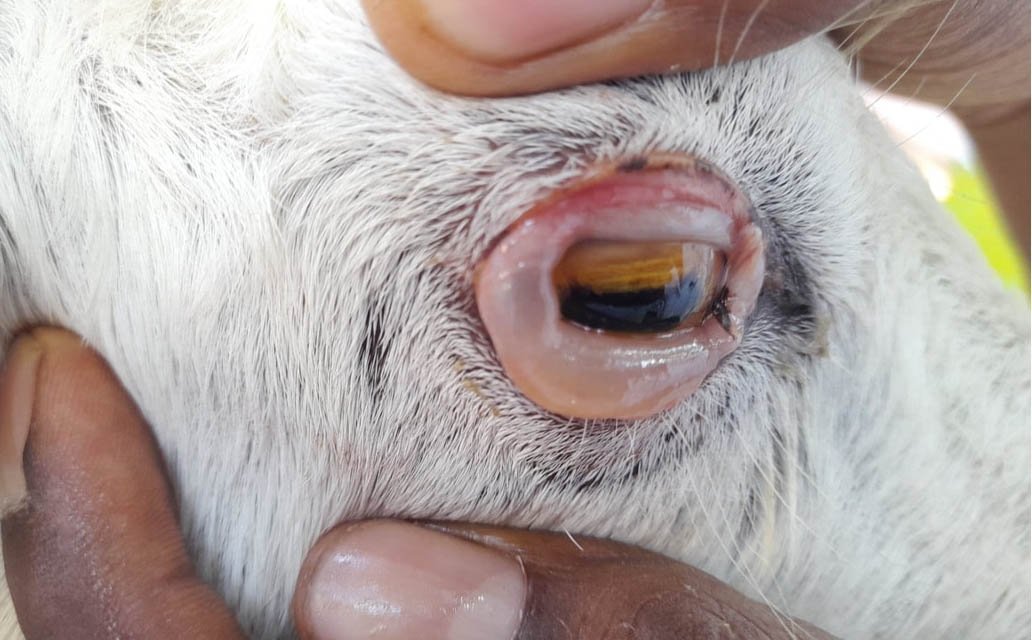
The normal appearance of the conjunctiva is pink, smooth and moist.
In systemic diseases, the appearance of the conjunctiva is altered. In cases of gastrointestinal disorders, it is congested; in jaundice, it is yellow; and it shows petechial (pinpoint haemorrhages) in toxaemia and septicaemia.
Echymosis of conjunctiva is noticed in protozon diseases like surra. It is dry and pale in shock, pale and watery in anaemia, ulcerated in riboflavin deficiency, and thickened in vitamin A deficiency (Xerophthalmia).
Etiology of Conjunctivitis
Bacterial or virus infection, Irritation due to chemical substances, Presence of foreign bodies, Trauma, Allergy and Nutritional deficiencies.
Classification
Based on etiology, conjunctivitis may be classified as specific conjunctivitis (e.g., seen in pink eye in horses, distemper in dogs), and non – specific conjunctivitis.
Clinically conjunctivitis is classified into three types, ciz., acute, subacute and chronic conjunctivitis.
According to the nature of inflammation the following varieties of conjunctivitis are recoreded-
- Catarrhal conjunctivitis, e.g., conjunctivitis due to mild bacterial infection or trauma.
- Purulent (suppurative) conjunctivitis, e.g., conjunctivitis seen in pink eye of horses, distemper of dogs, etc.
- Diphtheritic conjunctivitis, e.g., croupous conjunctivitis seen in birds. Diphtheritic conjunctivitis seen in calves due to infection by fusiformis necroforus.
- Granular or follicular conjunctivitis, causing small follicular enlargements on the conjunctiva known as trachoma.
Symptoms of Conjunctivitis
In the beginning stages of conjunctivitis lacrimation is thin and watery. Later it becomes thicker and has a tendency to stick on to the edges of lids and cheek.
Photophobia and blepharospasm are not marked in simple conjunctivitis. If these symptoms are present extension of inflammation to the cornea should be suspected.
Discomfort for the animal/dog and Chemosis (swollen conjunctiva through the palpebral fissure).
Diagnosis
Diagnosis of Conjunctivitis is based on history and clinical examination. it can be easily diagnosed.
Treatment
The conjunctival sac is irrigated at frequent intervals with warm saline solution or a mild antiseptic lotion.
The eye lotions commonly used were: ZAB lotion (zinc sulphate 1⁄2 %, alum 1%, boric acid 2%), percholride of mercury lotion (1 in 30,000 to 1 in 10,000), argyrol (5%) and boric lotion (2 to 3 %).
5% povidone Iodine can be used to cleanse the eye, followed by topical antibiotics and NSAIDS if necessary. Always check the integrity of the cornea prior to use of a corticosteroid.
“Chloromycetin applicaps” are found effective in many cases of conjunctivitis due to bacterial infection. Other antibiotic eye ointments like “teramycin eye ointment” are also effective. Hydrocortisone eye ointments are indicated in allergic conjunctivitis.