TABLE OF CONTENTS
Otitis
Otitis in animals is a inflammatory condition of the ear due to infections.
Otitis can be classified in three types-
Ottis externa is the inflammation of the external auditory canal, Otitis media is the inflammation of the middle ear and Otitis interna is the inflammation of internal ear. these all are quite related to each other and widely known as otitis.
Untreated ottitis externa leads the infection to inside the ear and cause ottitis media, further it leads to otitis interna.
Otitis externa
Otitis externa is defined as inflammation of the external auditory canal or external ear.
Etiology
- Numerous and multifactorial
- Peculiar anatomy and presence of hair in the canal
- Infection – Staphylococcus, streptococcus, pseudomonus, proteus, E.coli and Corynebacterium
- Yeast and fungus
- Parasitic
- Atopy Food allergy
- Metabolic diseases- hypothyrodism
- Immnune mediated
- Keratinisation disorder
- Accumulation of ear wax
Pathophysiology
Inflammatory process causes damage to the superficial protective stratum corneum of the canal. Hyperplasia and hypertrophy of sebacious and ceruminous glands. Infections may occur secondary to inflammation caused by both bacteria (Staphylococcus intermedius and Pseudomonas aeruginosa ) and fungal (Malasseziapachydermatis) organism, as well as small organisms such as ear mites. Cellular infiltration of mast cells, macrophages, lymphocytes, plasma cells, neutrophils, esinophils, retention of wax, erosions and ulcerations occurs within the canal resulting in serum exudates and necrotic debris.
Clinical Signs
- Pruritus manifested by scraching , rubbing the ear and shaking,
- mild erythema of the ear canal,]
- pain on palpation exudation from the ear
- proliferative granulation tissue in chronic cases
- calcification of the ear cartilage
Diagnosis
- Clinical signs and physical examination
- Radiography
- Video otoscopy
- Cytological study
- Antibiogram (ABST)
- Allergy testing
Treatment
Examine the ear canal under sedation/ GA. Plug the ear canal, Clip the hairs around the ear canal, Collect sample of discharge/swab, Clean the ear canal using cotton swabs dipped in isopropyl (rubbing) alcohol, NS , boric acid and acetic acid (vinegar) in various proportions and certain recipes containing povidone-iodine (betadine) to remove debri or any byproducts of the inflammation (exudate) or infection (pus) which lead to further irritation and discomfort, and may be in turn, causes of further infection.
- Medical irrigation with 1 in 100 povidone Iodine (Avoid if typanum is ruptured)
- Specific antimicrobials both systemic and topical based on ABST
- Anti inflammatory drugs
- Application of agents to dissolve the ear wax
Surgical procedure: Zepp’s operation
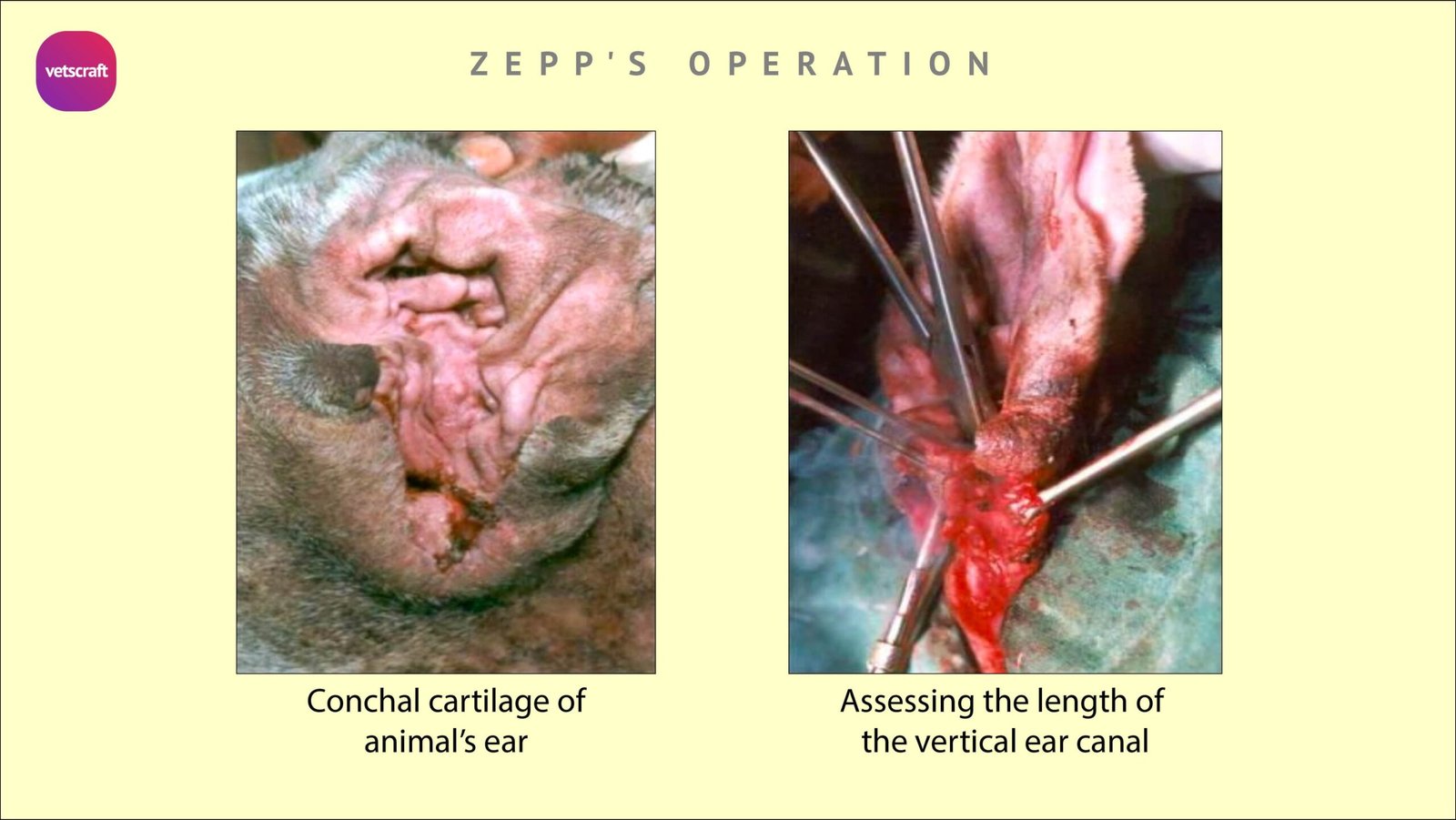
Site: Tubular antero external aspect of the concha
Two long curved forceps are applied on either side of the conchal cartilage with the apex of the V not connected.
Incisions are made on the conchal cartilage and the skin incision is reflected and the conchal cartilage is reflected down and bends to form a board. The drinage will be direct, Sutures are placed in a continous manner.
Otitis media
Otitis media is the inflammation of middle ear. it is common in dogs, goat, buffaloes and pigs. Inflammation may be initiated via three routes-
- Extension of otitis externa and Infection across the tympanic membrane through a rupture.
- Upper Respiratory Tract infection through Eustachian tube,
- Haematogenous spread: Trauma, Polyps, neoplasms and FB in the middle ear may also cause middle ear disease.
Pathophysiology
Swelling and occlusion of eustachian tube, Erosion of MM of tympanic cavity, Metaplasia of epithelium of ME, Degeneration or proliferative changes of tympanic bullae and accumulation of inflammatory debri in tympanic cavity.
Clinical signs
- General signs of otitis externa
- Facial Nerve Paralysis or Horness syndrome: due to affection of facial and sympathetic nerve since they pass near the middle ear. It may cause drooling of saliva, lack of palpebral reflex, ptosis, miosis, enophthalmos and protrusion of third eyelid.
- If the middle ear infection is chronic or severe enough to cause otitis interna, signs of abnormal vestibular functions (Nystagmus, ataxia, head tilt ) can occur.
Treatment
Medical treatment for Otitis media is MYRINGOTOMY (Incision on tympanic membrane ). It provide drainage from the ME cavity, provision for lavage and instillation of medicine and relieve pain and pressure. If the TM is discolored and bulged, collect the sample of fluid for ABST and Cytological studies.
Method is Clean the external ear canal. Introduce otoscope. A blunt probe is directed thr’ the otoscope cone to perforate the tympanic membrane caudal to malleus. Aspirate the fluid. Flush ME if needed by cannulation using NS until exudation is cleared. Systemic antibiotics should be advised.
Surgical Treatment
Surgical Treatments for Otitis media are-
- Lateral bulla osteotomy
- Ventral bulla osteotomy
Lateral bulla osteotomy
General anaesthesia –Lateral recumbency- Skin incision over Vertical canal extending 1-2 cm ventral to the horizontal canal. Dissect the s/c tissue to reveal the junction between the parotid gland and ventral aspect of the HC.
Further dissection along the ventral and caudolateral aspect of the HC exposes the facial nerve as it emerges from the stylomastoid foramen.
Retract the facial nerve ventrally to expose the lateral aspect of the bulla. The tissue overlying the bulla is incised and elevated with a periosteal elevator.
The bulla may be entered thr’ a steinmann pin or pneumatic drill. Extend the opening cranially, caudally and ventrally. The cavity is gently curetted and irrigated with Sterile NS.
Fix a temporary drain tube and secured to the bulla. The s/c and skin are closed, taking the drain tube out thr’ a skin incision 1-2 cm below and secure it to the skin.
The tympanic cavity is lavaged with an appropriate antiseptic / antibiotic as per the result of ABST. The drain tube may be removed after 7-10 days.
Ventral bulla osteotomy
This technique has the advantage of providing improved exposure and more consistent ventral drainage of the tympanic bulla than the lateral bulla osteotomy. Disadvantage is the technical difficulty.
General anaesthesia– dorsal recumbency- The area surrounding the angle of jaw is prepared for aseptic surgery. The bulla can be palpated immediately caudal and slightly medial to the vertical ramus of the mandible.
An 8-10 cm long paramedian incision is made just medial to the mandibular salivary gland and centered midway between the angular process of the mandible caudally and wings of the atlas cranially.
Incise the platysma muscle longitudinally for the length of the skin incision. Separate digastricus from hypoglossal and styloglosal muscles.
Deep digital palpation in this tissue plain confirm the location of the bulla as a raised rounded structure between the jugular process of the skull caudally and the angular process of the mandible rostrally.
Blunt dissection is continued till the bulla is reached.
The tympanic cavity may be entered using steinmann pin and the opening is widened. The interior of the bulla is examined for any FBs, inflammatory debri or neoplasm.
The cavity is irrigated and a bone curette is used to remove the remaining epithelial lining and drain tube may be fixed.The tube must exit thr’ the ventral cervical skin.
Otitis interna
Inflammation of the inner ear is called otitis interna, most often caused by an infection. The infectious agent is most commonly bacterial, although fungus can also be implicated in an inner ear infection in animals.