TABLE OF CONTENTS
Prolapsed nictitans gland
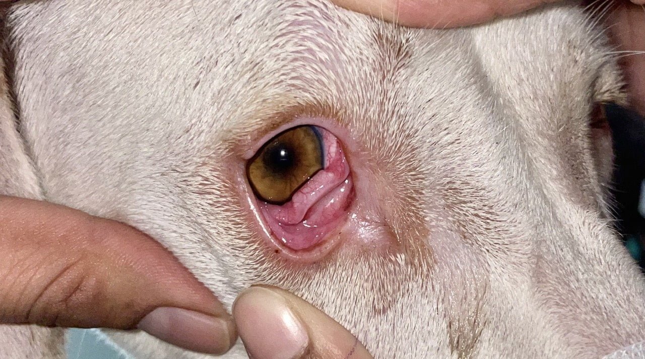
Prolapsed nictitans gland is the condition where the third eyelid prolapse in animals. This third eyelid prolapse is also known as cherry eye.
The resulting characteristic appearance of a pink, smooth-surfaced swelling protruding around the leading edge of the third eyelid from its inner surface.
The nictitans membrane gland of the third eyelid surrounds the base of the T-shaped cartilage-
- T-Cartilage forms the skeleton of the third eyelid
- This gland contributes to the aqueous and mucus phases of the precorneal tear film
- This is important in maintaining a healthy ocular surface
Dogs treated with surgical replacement of the gland have a lower incidence of dry eye later in life than dogs that are either left untreated or have the prolapsed gland excised.
Surgical procedure
Surgical procedure is the only treatment option for Prolapsed nictitans gland. there is two techniques are used-
- Orbital rim anchorage technique
- Conjunctival pocketing technique
Orbital rim anchorage technique
- The eyeball was fixed and it is flushed with a dilute Povidone Iodine
- An incision is made in the medio ventral conjunctival fornix using scissors.
- Blunt dissection allows access to the periosteum of the medioventral orbital rim.
- A firm bite of periosteum along the orbital rim is taken using 3 – 0 PDS.
- The needle is passed dorsally through the most prominent of prolapsed gland.
- Horizontal bite is taken through the dorsal prominence of the gland.
- Finally the needle is passed back through the last exit hole to emerge through the original incision thus encircling a large portion of the gland.
- The suture ends are then tied.
- Post operatively topical antibiotic is given.
Conjunctival pocketing technique
- The eyeball was fixed and it is flushed with a dilute Povidone Iodine
- An elliptical incision is made in the apex of the prolapsed gland with a No 11 BP blade and using scissors blunt dissection is performed and the conjunctival piece is dissected out. Further a pocket is made around the gland and the prolapsed gland is repositioned in to this pocket. Continous sutures are applied using 4-0 vicryl taking care to burry the suture ends.