TABLE OF CONTENTS
Amphistomes (Rumen Flukes): Morphology, Life Cycle, Pathogenesis, Diagnosis and Treatment
Amphistomes, commonly known as rumen flukes, are trematode parasites belonging to the family Paramphistomatidae. They primarily infect the rumen and reticulum of ruminants such as cattle, buffaloes, sheep, and goats. While adult flukes are generally non-pathogenic, immature stages migrating through the small intestine can cause severe enteritis and significant economic losses in livestock production.
Taxonomical Classification
- Kingdom: Animalia
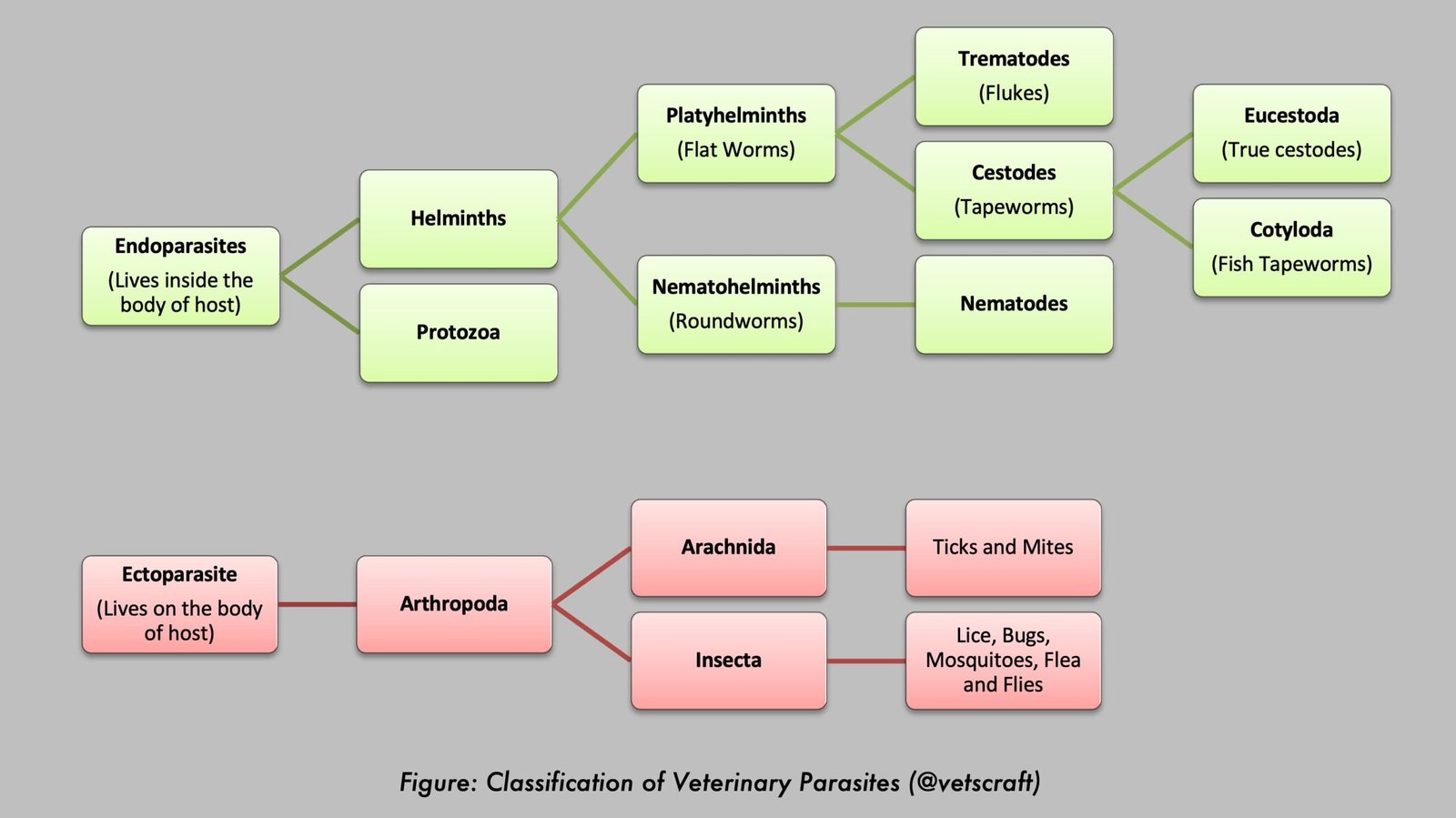
- Phylum: Platyhelminthes
- Class: Trematoda
- Subclass: Digenea
- Order: Echinostomida
- Suborder: Paramphistomata
Amphistomes
- Family: Paramphistomatidae
- Common Name: Rumen Flukes
- Disease: Immature Amphistomosis, Amphistomiasis
Important Amphistome Species Include:
- Paramphistomum cervi (P. gotoi)
- Gigantocotyle explanatum (Bile Duct Amphistome)
- Cotylophoron cotylophorum (Rumen Fluke)
- Gastrothylax crumenifer (Pouched Amphistome)
- Gastrodiscus secundus
- Gastrodiscus aegyptiacus
- Gastrodiscus hominis
- Pseudodiscus collinsi
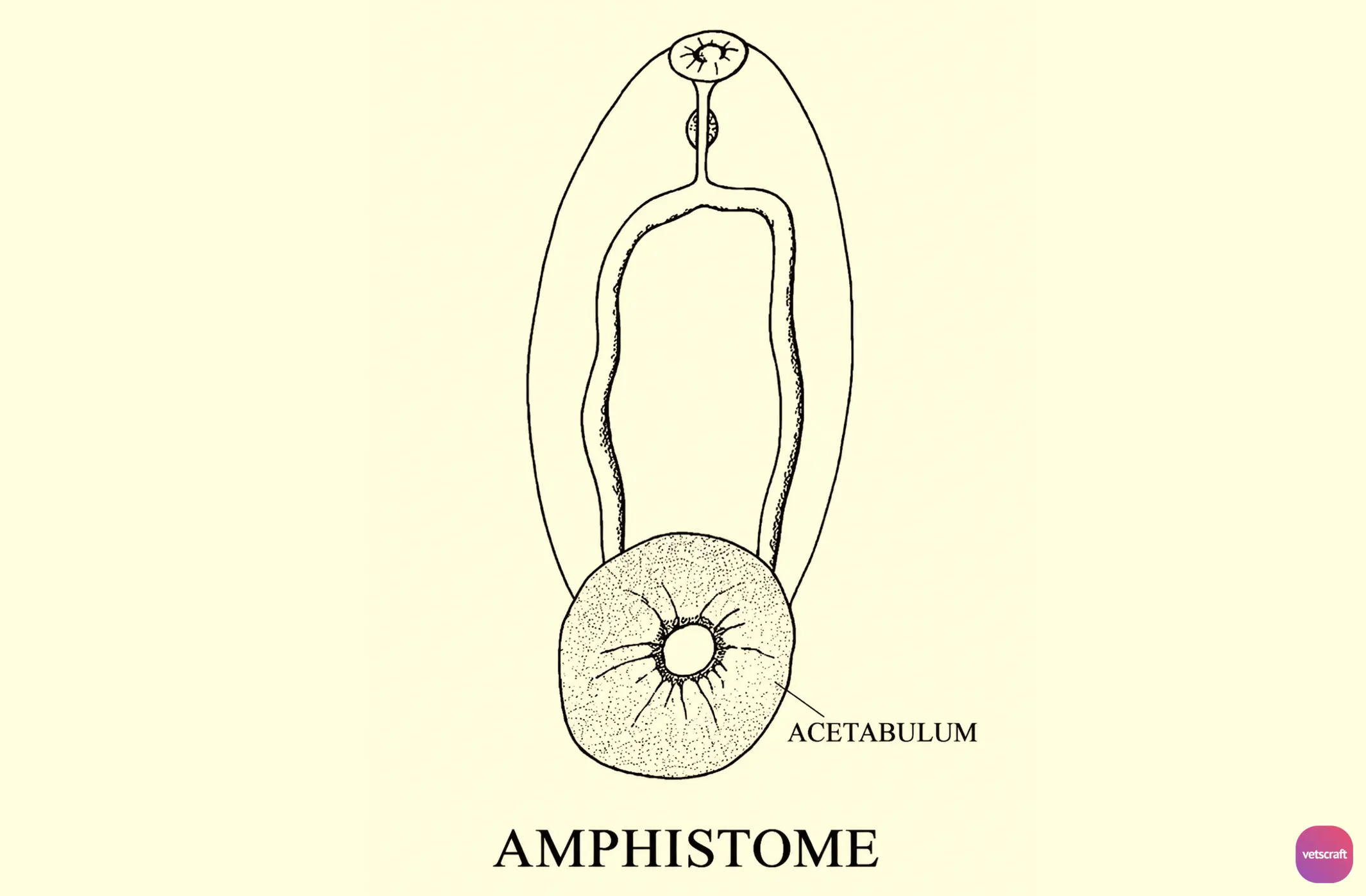
The terms amphistomes and rumen flukes are often used interchangeably, but they are not synonymous. Amphistomes are a diverse group of trematodes characterized by the presence of a posterior ventral sucker (acetabulum). While many amphistomes, such as Paramphistomum cervi, Cotylophoron cotylophorum, and Gastrothylax crumenifer, inhabit the rumen and reticulum of ruminants and are therefore known as rumen flukes, other amphistomes parasitize different organs. For example, Gigantocotyle explanatum occurs in the bile ducts, whereas species of Gastrodiscus and Pseudodiscus are intestinal parasites. Thus, all rumen flukes are amphistomes, but not all amphistomes are rumen flukes.
Characteristics of the Family Paramphistomatidae
- Flukes are fleshy, pear-shaped or conical in appearance (conical flukes) and are bright pale red in color when fresh.
- Spines are absent.
- The pharynx is absent, and the intestinal ceca are simple.
- The oral sucker (OS) is situated at the anterior end, and the ventral sucker (VS) is large and located at the posterior end.
- A ventral pouch may be present in some species, into which the genital ducts open.
- Testes may be arranged in tandem, diagonal, side-by-side, horizontal, or dorsoventral positions. The cirrus sac is absent.
- The ovary is situated posterior to the testes.
- Amphistome eggs are oval, operculated (with a distinct operculum), and possess a thin, transparent eggshell containing an early-stage embryo. The eggs have a small knob at the posterior extremity.
- All four larval stages occur during development. Snails act as intermediate hosts. The cercaria possesses eye spots and pigmentation and is therefore referred to as Cercaria pigmentata.
- Most amphistomes parasitize the gastrointestinal tract, except Gigantocotyle explanatum, which occurs in the bile ducts of the liver.
Life Cycle of Amphistomes
In amphistomes, eggs are passed in the feces of the host, and the miracidium develops within approximately 12–21 days.
The miracidium hatches from the egg and penetrates a freshwater snail.
Penetration of the snail occurs through the pneumostome and subsequently through the posterior wall of the mantle cavity. However, penetration through exposed soft tissues of the snail may also occur.
Young snails are more susceptible than older snails because the mantle cavity is completely filled with water and the pulmonary aperture remains permanently open.
The intermediate hosts of amphistomes are as follows:
- Indoplanorbis exustus: G. explanatum, C. cotylophorum, and G. secundus.
- Gyraulus convexiculus: G. crumenifer and G. explanatum.
- Lymnaea luteola: F. elongatus and F. cobboldi.
- Planorbis and Bulinus: Paramphistomum spp.
Following penetration of the snail, the miracidium loses its ciliated covering and transforms into a sporocyst. The sporocyst matures within 11 days, and each sporocyst contains approximately eight rediae.
Each redia produces 15–30 cercariae. Daughter rediae may occur under certain circumstances. Cercariae are released from the redia in an immature state and require approximately 13 days at 27°C to mature within the snail before being shed.
The mature cercaria is dark brown in color, possesses two distinct eye spots, and is pigmented; therefore, it is referred to as Cercaria pigmentata.
Cercariae are released from the snail during daylight hours. The released cercariae encyst on herbage or other objects in water. The metacercaria is dark in color and remains viable for up to three months.
Infection of the definitive host occurs through ingestion of metacercariae along with herbage. The ingested metacercariae excyst in the intestine.
The immature flukes attach to the mucosa of the duodenum.
Approximately 6–8 weeks after infection, the flukes migrate anteriorly through the reticulum and eventually reach the rumen. During migration, the flukes attach to the esophageal groove. The life cycle is completed within 3–4 months.
Epidemiology
In certain regions, immature amphistomosis is known by local names such as Pitto, Gillor, and Bisi.
Outbreaks of amphistomosis commonly occur during summer and dry seasons. During these periods, snail populations become concentrated around limited natural water sources where palatable grazing is also available. The availability of suitable grazing attracts large numbers of animals, resulting in the concentration of animals, snails, and metacercariae within a relatively small area. This situation predisposes animals to heavy infections.
Previous exposure and increasing age provide cattle with a degree of protection against reinfection. Consequently, acute or severe disease is more common in young animals than in adults. Older animals are generally capable of withstanding heavy exposure while continuing to contaminate pastures with eggs.
In contrast, sheep and goats remain relatively susceptible throughout their lives.
Pathogenesis of Rumen Flukes
Pathogenesis is primarily associated with immature amphistomes located in the duodenum and upper ileum during the intestinal phase of infection.
The immature stages become embedded in the mucosa and act as “plug feeders.” They draw portions of the mucosa into their suckers and pinch them off, resulting in necrosis and hemorrhage.
In severe infections, immature flukes become deeply embedded within the mucosa and may even extend into the muscular layer of the intestine, causing severe hemorrhagic duodenitis.
Adult amphistomes are generally non-pathogenic, even when present in large numbers. Their primary effect is the loss of ruminal papillae.
Histopathological Lesions
- Extensive catarrhal and hemorrhagic inflammation of the duodenum and jejunum, with destruction of the intestinal glands.
- Degeneration of associated lymph nodes and other organs.
Clinical Signs
In natural outbreaks, the predominant symptom is edema. It begins as a small, soft swelling in the subcutaneous tissue of the intermandibular space, which gradually increases in size and extends to the cheeks in sheep and to the dewlap and sternum in cattle, especially during the early morning and late evening. It is mainly caused by hypoalbuminemia. This condition is known as bottle jaw. In bullocks, edema may also develop in the subcutaneous tissue of the prepuce. The edema is soft on palpation.
Profuse, fluid, fetid diarrhea (“pea-soup diarrhea”) is the next predominant sign. Blood is usually absent, but the feces are dark in color and may contain strands of mucus as well as immature flukes. Chronic cases in cattle may result in rectal hemorrhage following a prolonged period of straining; in such cases, the feces may contain blood clots.
Appetite decreases and may eventually be completely lost. Affected animals generally show increased thirst. They drink small quantities of water frequently (polydipsia) and may keep their muzzle submerged in water for prolonged periods.
As diarrhea progresses, the animal develops signs of anemia, including very pale mucous membranes (almost white and bloodless), weakness, and prostration. Death may occur within 15 to 20 days after the onset of the first clinical signs.
A mortality rate of approximately 80–90% has been reported in acute outbreaks.
In some regions, immature amphistomosis is locally known by different names; however, these terms are not commonly used in international veterinary literature.
Postmortem Lesions
- Mucous membranes are pale, anemic, edematous, and the carcass is emaciated.
- Subcutaneous fat is replaced by gelatinous material.
- Hydrothorax and hydropericardium.
- Hemorrhagic duodenitis.
- Immature flukes are attached to the duodenal mucosa in clusters and appear pale pink in color.
Diagnosis
The diagnosis of the disease is based on:
- Clinical signs, usually observed in young animals.
- A history of grazing in areas with snail habitats during periods of dry weather.
- Fecal examination for eggs is of limited value because the disease occurs during the prepatent period when only immature flukes are present.
- Confirmation can be obtained through postmortem examination and recovery of small immature flukes from duodenal mucosal scrapings, or by demonstrating immature flukes in diarrheic feces.
- Collect approximately 500 mL of diarrheic feces from the affected animal, pass it through a fine-mesh sieve (approximately 0.053 mm aperture), and examine the residue under a stereomicroscope for the presence of immature flukes.
Treatment
- Niclosamide: 90 mg/kg body weight — effective against immature flukes.
- Oxyclozanide: 15–20 mg/kg body weight orally — effective against adult flukes.
- Triclabendazole: 10 mg/kg body weight.
- Niclofolan: 6 mg/kg body weight.
- Bithionol Sulfoxide: 40 mg/kg body weight.
- Resorantel: 65 mg/kg body weight.
Control
- Control of the snail intermediate host.
- Animals should be allowed to graze on higher ground whenever possible.
- Localized water bodies should be fenced off or treated with molluscicides. Proper drainage of ponds, pools, and swampy areas should be maintained.
- Regular strategic treatment during high-risk seasons.