
Histomonas meleagridis: Morphology, Life Cycle, Pathogenesis, Clinical Signs & Treatment
Histomonas meleagridis is a flagellated protozoan parasite that causes histomoniasis (blackhead disease), a highly important disease of turkeys and other gallinaceous birds. The parasite primarily affects the cecum and liver, where it produces characteristic necrotic lesions that can result in severe economic losses due to high mortality, poor growth, and reduced productivity. Although turkeys are the most susceptible species, chickens often act as reservoir hosts and play a major role in the epidemiology of the disease.
In this article, we’ll learn about the taxonomy, morphology, life cycle, transmission, pathogenesis, clinical signs, diagnosis, treatment, and control of Histomonas meleagridis in a concise, veterinary parasitology focused format.
Parasite Overview
- Species: Histomonas meleagridis
- Host: Turkeys; also occurs in chickens, peafowl, guinea fowl, Japanese quail, and partridges. Chickens act as reservoir hosts.
- Predilection Site: Cecum and liver
- Disease: Histomoniasis, infectious enterohepatitis, and blackhead disease
- Distribution: Worldwide
Taxonomical Classification
- Kingdom: Protista
- Phylum: Metamonada
- Class: Parabasalia
- Order: Trichomonadida
- Family: Monocercomonadidae
- Genus: Histomonas
- Species: Histomonas meleagridis
- Common Name: Blackhead disease protozoan, histomonad
Morphology
The body of Histomonas meleagridis is actively amoeboid, usually rounded but sometimes elongated, with a single nucleus and a flagellum arising from a basal granule located close to the nucleus.
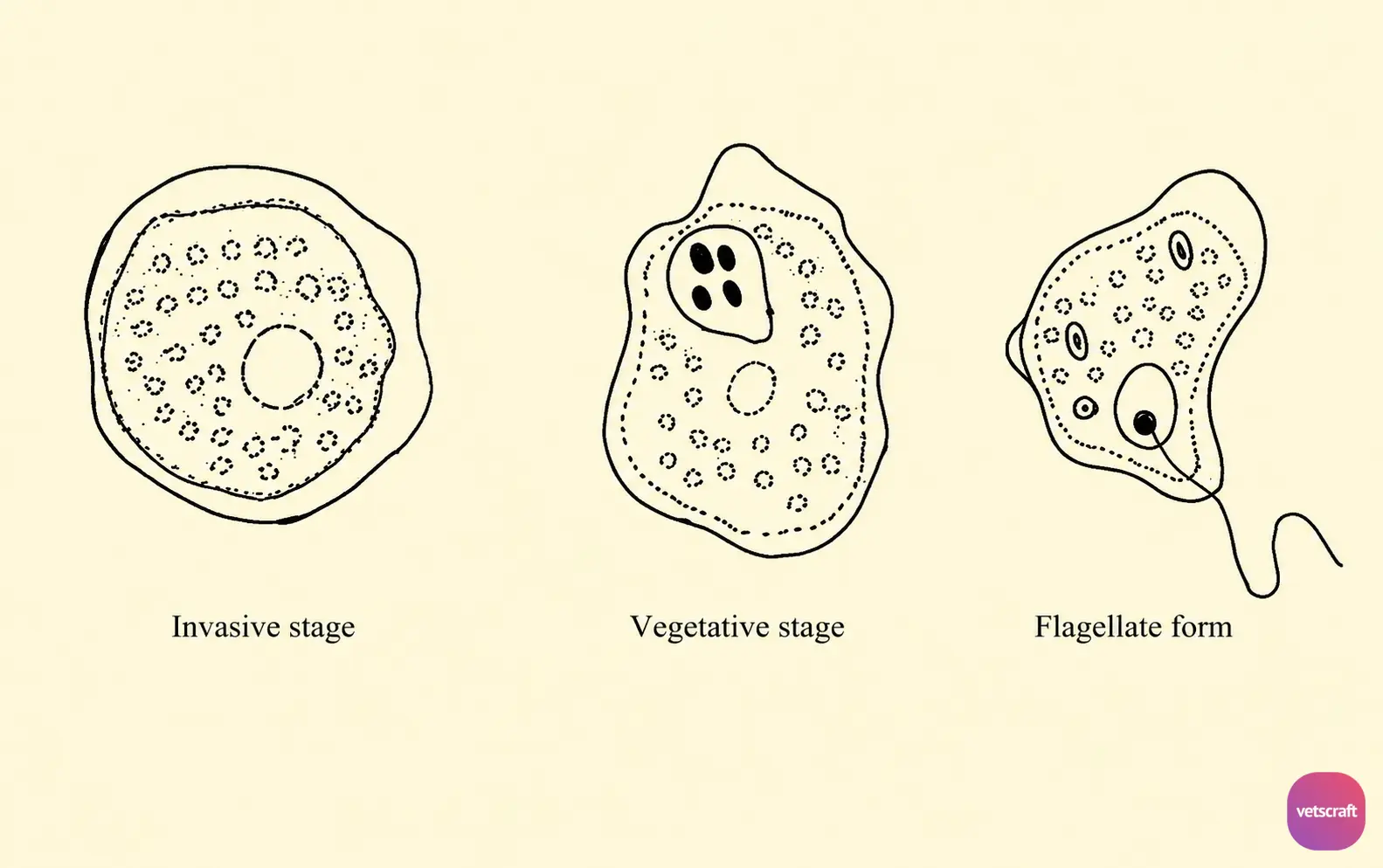
H. meleagridis is pleomorphic; therefore, its morphology depends on its location and the stage of the disease. Based on its structure and location in the host, four stages have been described in H. meleagridis.
The cytoplasm is divided into a clear ectoplasm and a granular endoplasm. Reproduction occurs by binary fission, and there is no evidence of a sexual cycle.
Different Stages of Histomonas meleagridis
- Invasive Stage is usually found in early cecal and liver lesions and at the periphery of older lesions. It is an extracellular, actively amoeboid stage with blunt, rounded pseudopods. The food vacuoles contain particles of ingested material but no bacteria.
- Vegetative Stage is found near the center of slightly older lesions. It is less active than the invasive stage and is packed tightly together, causing disruption of the tissues.
- Resistant Stage is compact, enclosed within a dense membrane, and found either singly or in clusters. It is usually extracellular but may be taken up by phagocytes or giant cells. Originally, it is no more resistant than the other stages.
- Flagellate Form is amoeboid in shape and occurs in the lumen of the cecum and in culture. It has a single vesicular nucleus and a flagellum. The flagellum produces characteristic jerky, oscillating movements resembling those of trichomonads. The endoplasm of this stage contains bacteria, starch granules, food particles, and erythrocytes. No cyst is formed.
Transmission and Life Cycle
Transmission of Histomonas meleagridis occurs in three ways:
1. Direct Transmission through Ingestion of Trophozoites
Turkeys can become infected by ingesting trophozoites, but this mode of transmission is uncommon because naked trophozoites are delicate and do not survive long in the environment. However, it may occur in flocks with an existing infection.
2. Transmission through Infected Heterakis gallinarum Eggs
The most important mode of transmission is the ingestion of Heterakis gallinarum eggs infected with H. meleagridis (an example of hyperparasitism). When these two parasites coexist in the cecum, H. meleagridis invades the developing eggs.
The infected eggs are eventually passed in the feces and undergo embryonation in the environment. During embryonation, H. meleagridis also multiplies rapidly and invades the larval nematode. Infected eggs can survive in the soil for at least 2 years.
3. Transmission through Ingestion of Earthworms (Transport Host)
If an earthworm ingests infected H. gallinarum eggs, the larvae hatch and remain infective for several weeks.
Development of the Organism in the Definitive Host
When a turkey ingests infected eggs or an infected earthworm, the larvae hatch in the intestine. The hatched larvae migrate to the cecum, their site of predilection, where Histomonas organisms are released from the larvae.
After being released, the organisms invade the cecal wall, multiply, and produce characteristic lesions. They may also be carried to the liver via the bloodstream, where they produce lesions.
Pathogenesis
Histomoniasis can affect turkeys of all ages, but the course of the disease and mortality vary with age. Poults less than 3 weeks of age are resistant to the disease, whereas those between 3 and 12 weeks of age are highly susceptible.
In this age group, the disease usually presents as an acute form and may cause 50–100% mortality. Affected birds typically die within 2–3 days after the onset of clinical signs. In older birds, the disease is generally more chronic, and recovery may occur. The mortality rate decreases as the age of the birds increases.
Chickens are less susceptible than turkeys, but severe outbreaks may occur in young birds. Histomonas is extremely common in Heterakis-infected chickens, which are the most important reservoir hosts of H. meleagridis. These birds serve as a source of infection for turkeys, either directly or indirectly through earthworms.
Clinical Signs
The incubation period is 15–21 days. The first sign of the disease is droopiness. Affected birds appear weak and drowsy and stand with a lowered head, ruffled feathers, and drooping wings and tail. They also develop sulfur-colored diarrhea. The head may or may not become darkened.
In histomoniasis, lesions are primarily observed in the cecum and liver. One or both ceca may be affected. Initially, the lesions appear as small, raised pinpoint ulcers containing numerous parasites. As the disease progresses, the lesions enlarge, and the entire cecum may become involved. Consequently, the cecal mucosa becomes thickened and necrotic and is covered with a characteristic foul-smelling yellowish exudate.
The exudate frequently becomes consolidated, leading to the formation of a dry, hard, cheesy plug that is firmly attached to the cecal wall. The ceca become markedly inflamed. In some cases, the ulcers may perforate the cecal wall, resulting in peritonitis.
The liver lesions are pathognomonic and appear as circular, depressed, yellowish to yellowish-green areas of necrosis and tissue degeneration. The lesions may reach up to 1 cm in diameter and extend deep into the liver. In older birds, the lesions are often confluent. Other organs, such as the kidneys and lungs, may occasionally be affected.
If the birds recover, the parasites disappear from the tissues, and the repair process begins. The cecal plug contracts, becomes smaller, and is eventually passed out. In less severe cases, the ceca may appear normal, whereas in severe cases, extensive scarring of the cecum and liver may persist for a long time.
Immunity
Birds that recover from histomoniasis develop immunity to reinfection.
Diagnosis
- Based on the clinical signs.
- Based on characteristic lesions in the liver and cecum.
- Histopathological examination of the lesions reveals hyperemia, hemorrhage, lymphocytic infiltration, and necrosis.
Treatment
- Enheptin can be administered through feed at a concentration of 0.05% for prevention and 0.1% for treatment.
- Furazolidone: 0.01–0.02% in feed.
- Dimetridazole: 0.0125% in feed.
Control
- Avoid contact between turkeys and domestic chickens.
- Turkeys and chickens should not be housed on the same premises.
- The use of hens for incubating and rearing turkey poults should be avoided.
- Young birds should be raised in cage systems, and droppings should be removed regularly.
- Young turkeys should be kept separate from adult birds.

