
Entamoeba histolytica (Amoebiasis): Morphology, Life Cycle & Pathogenesis
Entamoeba histolytica is a pathogenic amoeboid protozoan that primarily infects the large intestine of humans and can occasionally affect several animal species. It is the causative agent of amoebiasis (amoebic dysentery), a disease characterized by intestinal inflammation and, in severe cases, extraintestinal involvement of organs such as the liver, lungs, and brain. The parasite is widely distributed worldwide and is transmitted through the ingestion of infective cysts in contaminated food or water.
E. histolytica has a direct life cycle consisting of trophozoite and cyst stages, with the mature quadrinucleate cyst serving as the infective stage.
Parasite Overview
- Host: Humans, monkeys, dogs, cats, pigs, and cattle
- Predilection Site: Large intestine; occasionally, it may also be found in the lungs, brain, and spinal cord.
- Disease Caused: Amoebic dysentery (Amoebiasis)
- Distribution: Worldwide
Taxonomical Classification
- Kingdom: Protista
- Phylum: Amoebozoa
- Class: Archamoebae
- Order: Entamoebida
- Family: Entamoebidae
- Genus: Entamoeba
- Species: Entamoeba histolytica
Morphology
Two developmental stages, the trophozoite (vegetative, motile stage) and cyst (resistant stage), are found in E. histolytica. Reproduction (multiplication) occurs by binary fission.
Trophozoite
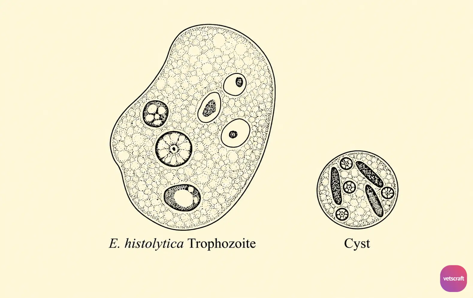
The trophozoite has a thick, clear ectoplasm and granular endoplasm. It is an actively motile stage that moves rapidly forward in a straight line with a single pseudopodium at the anterior end.
The pseudopodium appears suddenly as a long, finger-like projection, followed by the flow of endoplasm into it. It multiplies by binary fission.
There is a single spherical nucleus containing a central endosome surrounded by a clear zone or halo. The nuclear membrane is lined with fine chromatin granules, giving the appearance that the nucleus is outlined by a ring of small beads. The nucleus is indistinct in living specimens.
The active trophozoite possesses food vacuoles that contain erythrocytes. The presence of erythrocytes in the trophozoite stage is an important characteristic of E. histolytica, which differentiates it from non-pathogenic forms.
Cyst
The cyst is spherical or ovoid in shape and measures 5–20 µm in diameter. The cyst wall is visible in living specimens but not in stained specimens. Initially, the cyst is uninucleate but becomes quadrinucleate upon maturation.
The mature quadrinucleate cyst is the infective stage of the parasite. It contains chromatoid bodies and glycogen.
The chromatoid bodies appear as rods with rounded ends, and the glycogen vacuoles are clearly visible in young cysts but disappear as the cyst matures.
Epidemiology
E. histolytica is primarily a parasite of humans. Since animals acquire infection from humans through direct contact, the transmission is known as zooanthroponosis. Infection in humans and animals occurs through the ingestion of quadrinucleate cysts in contaminated water, food, or raw vegetables.
Houseflies play an important role in contaminating food with amoebic cysts. The cysts are relatively resistant and are not affected by water chlorination but can be removed by sand filtration.
Faulty plumbing and water systems may result in waterborne transmission. Food handlers may also play an important role in the transmission of amoebiasis. The cysts do not survive for more than 10 minutes on the hands.
Life Cycle
When mature quadrinucleate cysts are ingested by humans or animals, they pass through the stomach and undergo excystation in the cecum or the lower part of the ileum.
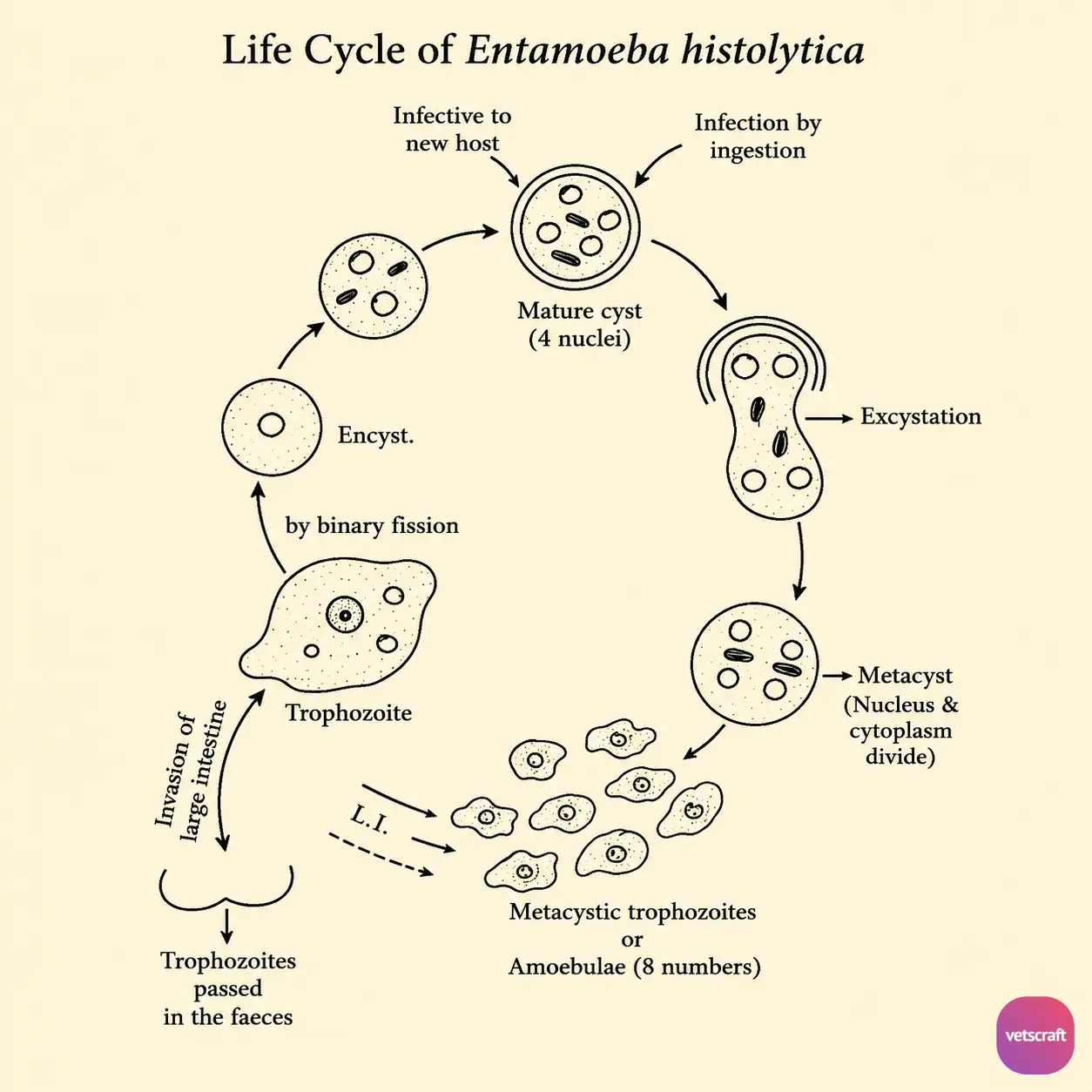
Following excystation, an amoeba with four nuclei, called a metacyst, is liberated from each cyst. This metacyst undergoes division to produce eight small uninucleate amoebulae (metacystic trophozoites). These young amoebulae then invade the submucosal tissue of the large intestine, where they develop into large trophozoites.
The large trophozoites multiply by binary fission and form colonies in the cecum and colon. These active, motile trophozoites are responsible for the pathogenesis and lesions associated with amoebiasis.
Once the trophozoites enter the distal portion of the large intestine (colon), they begin to disgorge undigested food material and condense into a spherical mass. This stage, known as the precyst, marks the beginning of cyst formation. The young cyst initially contains a single nucleus with a thin wall, but after two rounds of nuclear division, it becomes a binucleate and then a quadrinucleate cyst.
As the cyst matures, the glycogen vacuoles disappear, while chromatoid bodies are formed. The mature cyst develops a thick wall and contains four nuclei and chromatoid bodies. These mature quadrinucleate cysts are passed in the feces and are ready to infect a susceptible host.
Pathogenesis
The large trophozoites are responsible for the pathogenesis and lesions associated with amoebiasis.
Based on the location of the lesions, the pathogenesis of E. histolytica is divided into:
- Primary or Intestinal Amoebiasis: In this form, the infection is limited to the large intestine and is usually manifested as acute amoebic dysentery or chronic intestinal amoebiasis.
- Secondary or Extraintestinal Amoebiasis: This extraintestinal form occurs due to the migration of trophozoites to various organs such as the liver, lungs, and brain.

