
Leishmania Parasite: Developmental Stages, Transmission & Species
Leishmania is a genus of protozoan hemoflagellate parasites belonging to the family Trypanosomatidae. These parasites infect humans, domestic animals, and wildlife and are transmitted through the bite of infected female sand flies. Leishmania species are of significant medical and veterinary importance because they cause leishmaniasis, a group of diseases with cutaneous, mucocutaneous, and visceral forms.
The life cycle of Leishmania alternates between a vertebrate host and a sand fly vector, involving two distinct developmental stages: the intracellular amastigote and the extracellular promastigote.
In 1900, Sir William Leishman first discovered Leishmania spp. in spleen smears from Dum Dum, India. Three years later, in 1903, Charles Donovan recorded the same organism in Chennai. Since this parasite was identified by two scientists independently within a very short period, it was named Leishmania donovani in their honor.
Members of this genus are obligate intracellular parasites of vertebrate hosts and are found within macrophages. They are heteroxenous parasites transmitted by sand flies (Phlebotomus spp.) in India and Lutzomyia spp. in other countries. Transmission occurs through inoculation when infected sand flies feed on susceptible hosts.
Developmental Stages of Leishmania
Only two developmental stages, amastigote and promastigote, are present in Leishmania spp.
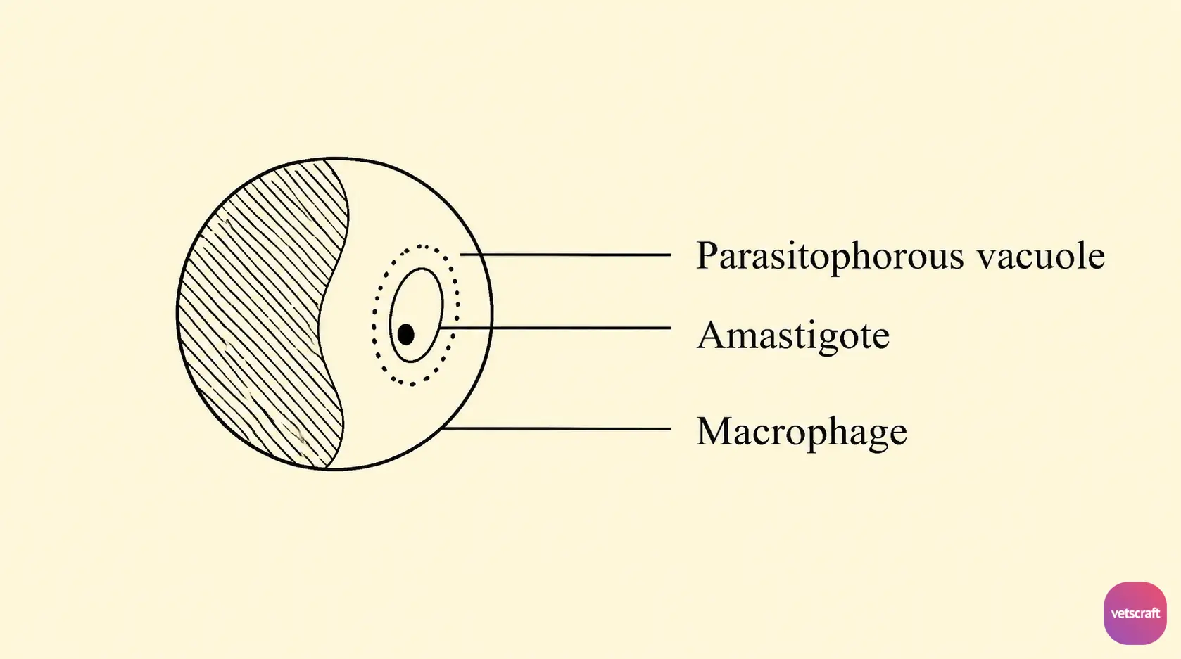
The amastigote is ovoid or spherical in shape (2.5–5 µm) and non-motile. The nucleus is located on one side, while the kinetoplast is positioned at a right angle to it. The flagellum is small or degenerated; hence, it is referred to as a micromastigote. It is usually found in the macrophages and reticuloendothelial cells (spleen, liver, bone marrow, lymph nodes, mucosa, and skin) of the vertebrate host, where it multiplies by binary fission.
Within the cytoplasm of macrophages, the amastigote stage is found in clusters, usually within parasitophorous vacuoles. Amastigotes found in the bloodstream (monocytes and leukocytes) are called Leishman-Donovan bodies.
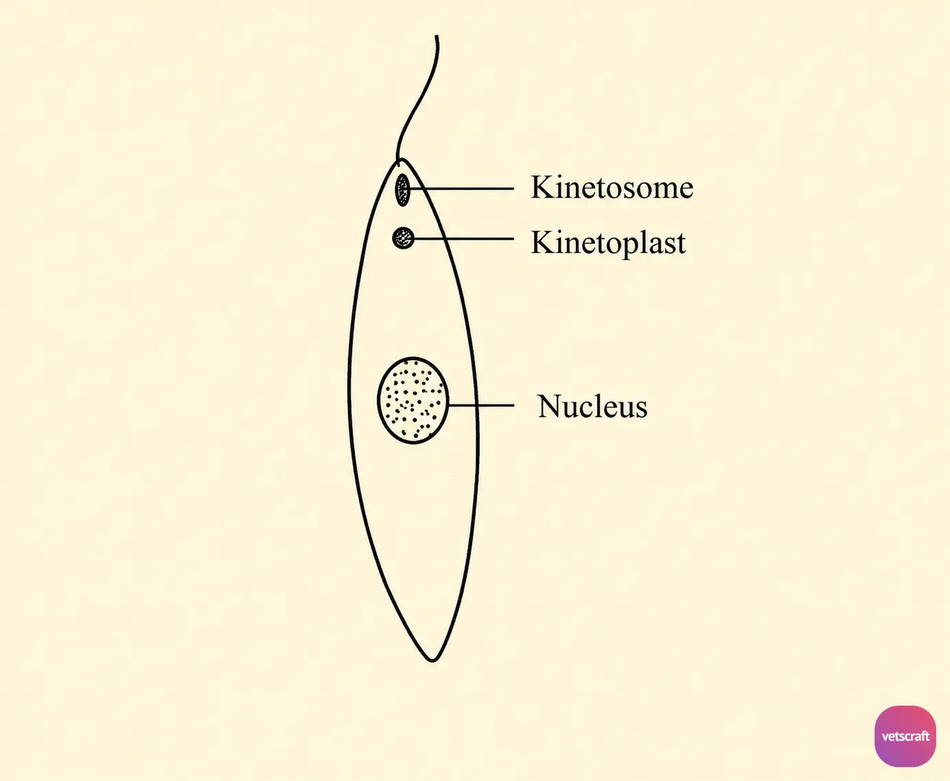
The promastigote is spindle-shaped (14–20 µm) and motile. It has a free flagellum as long as the body but lacks an undulating membrane. This is the infective stage of Leishmania, found in the gut of the insect vector.
Development and Transmission of Leishmania
Development in the Vector (Phlebotomus)
When sand flies feed on an infected human or animal, they ingest leukocytes and macrophages containing amastigotes along with the blood meal.
The ingested amastigotes pass into the midgut of the insect, where they transform into promastigotes. These promastigotes multiply by binary fission, eventually producing large numbers of organisms.
These promastigotes then migrate to the esophagus and pharynx of the fly, where they accumulate, leading to blockage of the food canal. When an infected fly attempts to feed on a susceptible host, a plug of organisms may be dislodged and injected into the host.
Development in the Vertebrate Host
The inoculated promastigotes soon transform into the amastigote stage. In the vertebrate host, the amastigote stage of Leishmania is found within macrophages, the reticuloendothelial (RE) system, and leukocytes, especially large mononuclear cells.
Important Species of Leishmania and Associated Diseases
| Name of the Species/Subspecies | Name of the Disease |
|---|---|
| Leishmania donovani | Kala-azar (visceral leishmaniasis), black fever, and Dum Dum fever |
| Leishmania tropica | Cutaneous leishmaniasis (Delhi boil, Oriental sore) |
| Leishmania tropica subsp. major | Wet (rural) cutaneous leishmaniasis |
| Leishmania tropica subsp. minor | Dry (urban) cutaneous leishmaniasis |
| Leishmania chagasi | New World cutaneous leishmaniasis |
| Leishmania mexicana | Cutaneous and mucocutaneous leishmaniasis |
| Leishmania braziliensis | Espundia (mucocutaneous leishmaniasis) |
| Leishmania peruviana | Uta |

