
Trypanosoma evansi (Surra): Classification, Morphology, Pathogenesis, Diagnosis & Treatment
Trypanosoma evansi is a pathogenic hemoflagellate protozoan that causes trypanosomosis (surra), a major vector-borne disease affecting domestic and wild animals worldwide. It is mechanically transmitted by biting flies and primarily infects horses, camels, cattle, buffaloes, dogs, and several other mammalian species. The disease is characterized by intermittent fever, anemia, edema, progressive weight loss, reduced productivity, and, in severe cases, death if left untreated.
This comprehensive guide covers the taxonomy, morphology, epidemiology, transmission, pathogenesis, clinical signs in different animal species, diagnosis, treatment, and control of Trypanosoma evansi. It is designed to serve as a reliable study resource for veterinary students, competitive examination preparation, and clinical veterinary practice.
Trypanosoma evansi was the first pathogenic trypanosome, recorded by Griffith Evans in 1881 in India. The disease caused by Trypanosoma evansi is known as trypanosomosis.
Parasite Overview
- Host: It affects a wide range of animals, including horses, camels, cattle, water buffalo, swine, donkeys, dogs, elephants, goats, tigers, and leopards.
- Location: Blood and lymph as an extracellular parasite.
- Vector: Tabanus spp. and other biting flies such as Stomoxys, Chrysops, and Haematopota spp.
The following terms are used for this disease in different animal species:
- Horse: Surra (Hindi term meaning “rotten”)
- Camel: Tibersa (referring to its chronic course of approximately 3 years), also known as Gufar
- Elephant: Thut
Taxonomical Classification
- Kingdom: Protista
- Phylum: Euglenozoa
- Class: Kinetoplastea
- Order: Trypanosomatida
- Family: Trypanosomatidae
- Genus: Trypanosoma
- Subgenus: Trypanozoon
- Species: Trypanosoma evansi
Epidemiology
Trypanosoma evansi is more prevalent in tropical and subtropical regions of the world. It is one of the most important trypanosomes affecting domestic and wild animals. The occurrence of surra is usually high during the rainy season (June–November) because vector populations increase during this period.
Camels, cattle, and sheep act as carrier hosts from which the infection is transmitted to susceptible hosts. Although cattle often serve as carrier hosts, they may develop clinical signs when subjected to stressors such as vaccination, transportation, intercurrent infections, parturition, or other stressful conditions.
Donkeys and mules are more resistant to trypanosomosis.
Morphology
- The body is leaf-like, fusiform, and measures 15–24 µm in length.
- It is usually monomorphic; however, polymorphic forms such as slender, intermediate, or stumpy forms may occasionally occur.
- The nucleus is vesicular and contains an endosome.
- A single flagellum arises from the kinetosome (basal granule, blepharoplast, or parabasal body), which is situated at the posterior end. An elongated blind pouch near the kinetosome is known as the flagellar pouch. It possesses a well-developed undulating membrane and a substantial free flagellum (5–6 µm) at the anterior end.
- The kinetoplast is located subterminally, posterior to the kinetosome. It is rod-shaped or spherical and contains DNA.
- The posterior end is not pointed (i.e., it is rounded or truncated).
Transmission
In herbivores, T. evansi is transmitted mechanically by biting flies such as Tabanus spp., Stomoxys spp., Chrysops spp., and Haematopota spp. It may also be transmitted through contaminated needles, which is referred to as iatrogenic transmission.
Among the various biting flies, the intermittent feeding habit of Tabanus facilitates the rapid transmission of T. evansi from one host to another. Therefore, it is considered a highly efficient mechanical vector.
In carnivores (dogs, cats, lions, and tigers), T. evansi is transmitted through ingestion of infected meat (carnivorism). The trypanosomes may enter the body through mucosal abrasions or wounds caused by bone fragments.
In the vertebrate host, T. evansi multiplies by longitudinal binary fission in the bloodstream trypomastigote stage.
Pathogenesis
The degree of pathogenicity depends on the virulence of T. evansi and the host species involved.
The pathogenic effects of trypanosomes include:
- Anemia
- Edema
- Metabolites released by rapidly multiplying trypanosomes disrupt normal physiological processes and cause tissue degeneration.
- Host tissue proteins are degraded by the excretory and secretory proteases of trypanosomes.
- Because trypanosomes have a high rate of glucose metabolism, they compete with the host for glucose, leading to hypoglycemia. This may be one of the causes of death in trypanosomosis.
- Salivarian trypanosomes possess a unique characteristic known as antigenic variation (variant surface glycoproteins), achieved by frequent changes or shedding of their surface coat. Consequently, the host’s immune system becomes exhausted due to the continuous production of large quantities of immunoglobulins against successive antigenic variants, resulting in hypergammaglobulinemia.
- Polyclonal activation of immune cells followed by marked immunosuppression may predispose affected animals to secondary bacterial infections.
Reason for Anemia in Trypanosomosis
- Flagellar movement causes mechanical destruction of RBCs.
- Immune-mediated erythrophagocytosis: Attachment of trypanosome antigens and adsorption of Ag–Ab complexes onto the surface of RBCs may predispose them to erythrophagocytosis.
- Enzyme-mediated erythrophagocytosis: Sialic acid present in the RBC membrane is degraded by the enzyme sialidase produced by trypanosomes. RBCs lacking sialic acid are more susceptible to erythrophagocytosis.
- Extravascular destruction of RBCs.
- Hemolytic factors cause direct hemolysis of RBCs.
- Coagulation disorders, including thrombocytopenia and intravascular coagulation.
Reason for Occurrence of Edema
The Ag–Ab complexes formed during infection are activated by Hageman factor. The activated Ag–Ab complexes, in turn, activate the kininogenase precursor, prekallikrein, resulting in the activation of kininogens and subsequent release of kinins. These kinins increase vascular endothelial permeability, leading to edema.
Causes of Death in Trypanosomosis
- Anemia
- Hypoglycemia
- Acidosis resulting from increased production and accumulation of lactic acid.
- Reduced oxygen-carrying capacity of RBCs, leading to asphyxiation and death.
- Increased serum potassium concentration, causing cardiac depression.
Clinical Signs
Although the clinical signs of trypanosomosis vary among animal species, some signs are common to all affected animals.
These include:
- High intermittent fever (40–44°C).
- Anemia/jaundice.
- Edema of the ventral region and dependent parts.
- Lymphadenitis.
- Emaciation/cachexia.
- Nervous disorders.
The course of the disease is categorized into two phases: paroxysms, during which parasites are present in the circulation along with fever, and intermissions, during which parasites are absent from the circulation, with or without fever.
Clinical Signs in Horses
“Surra” is a Hindi term meaning “rotten,” referring to a condition of severe or progressive wasting (chronic wasting disease).
Trypanosomosis is almost always fatal in horses if left untreated. Death may occur within a few weeks to 6 months, depending on the strain and virulence of the organism. The disease may follow either an acute or chronic course.
Acute Form
The incubation period is 4–9 days. Clinical signs commonly observed in the acute form include intermittent fever, emaciation, and edema of the dependent parts, particularly the fetlocks, brisket, and preputial sheath in males.
Urticarial plaques may be present on the neck and flank regions. Hemorrhages occur at the mucocutaneous junctions, especially around the nostrils, eyes, and anus. Paralysis of the hindquarters (Mal de Caderas) may also occur.
Chronic Form
The chronic form is characterized by enlargement of the lymph nodes and hemorrhages in the anterior chamber of the eyes and vulva. If trypanosomes invade the CNS, affected animals may develop hindquarter weakness with dragging of the hooves and profuse sweating.
Postmortem Lesions in Horses
Anemia, emaciation, enlargement of lymph nodes, splenomegaly, and petechial hemorrhages on the serosal surfaces and within the parenchyma of the liver and kidneys.
Clinical Signs in Cattle and Buffalo
Cattle and buffalo are considered reservoirs of infection for equines. A subclinical form of the disease is common in bovines. However, an acute form with sudden death may occasionally occur. The disease is generally more severe in young and debilitated animals.
Based on the clinical manifestations, trypanosomosis in cattle is classified as:
Peracute Form
Animals may die within 2–3 hours after the onset of clinical signs. Before death, they may exhibit nervous signs such as convulsions. This sudden death may be mistaken for snakebite, anthrax, ketosis, milk fever, rabies, poisoning, or other acute conditions.
Acute Form
Animals become dull and sleepy, with the eyes remaining wide open (staring appearance due to nystagmus). Breathing becomes labored and noisy, accompanied by circling movements, staggering gait, and butting against walls or the manger.
Stamping of the feet, bellowing, teeth grinding, frequent urination, profuse salivation, muscle twitching, shivering, coma, and death may occur within 6–12 hours after the onset of clinical signs. Body temperature may rise to 39.4–40.6°C, although the fever is not always consistent.
Chronic/Subacute Form
The chronic form is characterized by dullness, lacrimation, emaciation, intermittent fever, edema of the legs, anemia, diarrhea, and eventual death.
Postmortem Findings in Cattle and Buffalo
Splenomegaly, hepatomegaly, enlargement of the kidneys and lymph nodes, and hemorrhages at the mucocutaneous junctions.
Clinical Signs in Camels
In camels, the disease is insidious and predominantly chronic. The course of the disease may last for up to 3 years; hence, it is called Tibersa in Asia and is also known as Gufar, in which death may occur after several months.
The chronic form is characterized by mild to severe congestion of the mucous membranes, edema, and disappearance of the hump due to fat mobilization.
An acute form may also occur, with an incubation period of 1–3 weeks. Affected animals exhibit weakness, loss of brightness of the eyes, dullness, anemia, alopecia, pallor, petechiae, edema of the legs, sheath, and dependent parts. Abortion is common, and mange infestation frequently accompanies trypanosomosis in camels.
Clinical Signs in Dogs and Cats
The disease is acute, fatal, and more severe in puppies than in adult dogs. The incubation period is 5–10 days.
Clinical signs include fever, anorexia, edema of the head, conjunctivitis, corneal opacity (pearl eye or white eye), blindness, and edema of the larynx, resulting in a change in voice (similar to that observed in rabies). In cats, corneal opacity may also be observed.
Clinical Signs in Sheep and Goats
Infected animals exhibit emaciation and anemia but usually do not show a loss of appetite.
Clinical Signs in Indian Elephants
The disease is commonly known as Thut. Affected elephants become markedly emaciated and exhibit muscular weakness, intermittent fever, dullness, listlessness, reluctance to move, anemia, and edema of the face and dependent parts.
Immunity in Trypanosoma evansi Infection
Persistence of immunity in recovered or carrier hosts is due to the continuous exposure of the host’s immune system to the parasites. Certain breeds, such as N’Dama, Muturu, and Brahman cattle, are relatively resistant to trypanosomosis and are referred to as trypanotolerant breeds. This resistance is mainly attributed to non-sterile immunity.
Trypanosomes, particularly those belonging to the salivarian group, possess a unique characteristic known as antigenic variation. They evade the host immune response by producing relapse strains with different antigenic characteristics through changes in their surface coat (variant surface antigens). As a result, regular fluctuations (parasitemia) occur in the number of organisms present in the blood and tissue fluids.
Antigenic variation in trypanosomes is one of the primary reasons why an effective vaccine against trypanosomosis has not yet been developed.
Immunosuppression
Severe immunosuppression occurs in trypanosomosis due to reduced B-cell production as well as the loss of memory cells.
Cultivation of Trypanosomes
Trypanosomes can be cultivated in a variety of media; however, NNN (Novy–McNeal–Nicolle) medium is the most commonly used. They can also be cultivated in developing chick embryos or in tissue culture media such as RPMI 1640 and DME supplemented with HEPES buffer.
Maintenance of Trypanosomes
Various laboratory animals are used for the maintenance of trypanosomes.
- In mice, trypanosomes can be maintained only for a very short period. The mice usually die within 3 days after inoculation.
- In rabbits, trypanosomes can be maintained for a longer period (3–5 months).
- In chickens, trypanosomes can also be maintained for an extended period.
Diagnosis
- Clinical signs.
- History and prevalence of tabanid vectors.
- Wet film examination: One of the best and quickest methods for diagnosing acute infection. A drop of blood is placed on a glass slide, covered with a coverslip, and examined under a microscope. Motile trypanosomes can be observed.
- Examination of thick and thin blood smears.
- Microhematocrit method: Used for the diagnosis of chronic or mild infections. In this technique, suspected blood is collected into a capillary tube, one end is sealed with wax or plasticine, and the tube is centrifuged for 5–10 minutes. After centrifugation, the capillary tube is placed on a glass slide and examined under a microscope. Motile trypanosomes can be observed at the plasma–buffy coat interface.
- Animal Inoculation Test: Laboratory animals such as mice, guinea pigs, and rabbits are used. Approximately 0.5 mL of suspected blood is inoculated intraperitoneally into mice or another laboratory animal. If the blood contains trypanosomes, death generally occurs within 2–3 days in mice, about 1 week in rabbits, and 7–8 days in guinea pigs.
- Chemical Tests: (Non-specific tests that may produce false-positive results.)
- Mercuric chloride test.
- Nitric acid test.
- Formol gel test.
- Stibamidine test.
- Thymol turbidity test.
- Other Tests:
- IFT, IHA, AGID, CIEP, CFT, ELISA, and agglutination or flocculation tests.
- DEAE-cellulose ion-exchange column.
The DEAE-cellulose ion-exchange column retains the more negatively charged erythrocytes while allowing the less negatively charged trypanosomes to pass through. This highly sensitive technique (Lanham, 1968) is used for harvesting trypanosomes from heavily infected blood.
Treatment
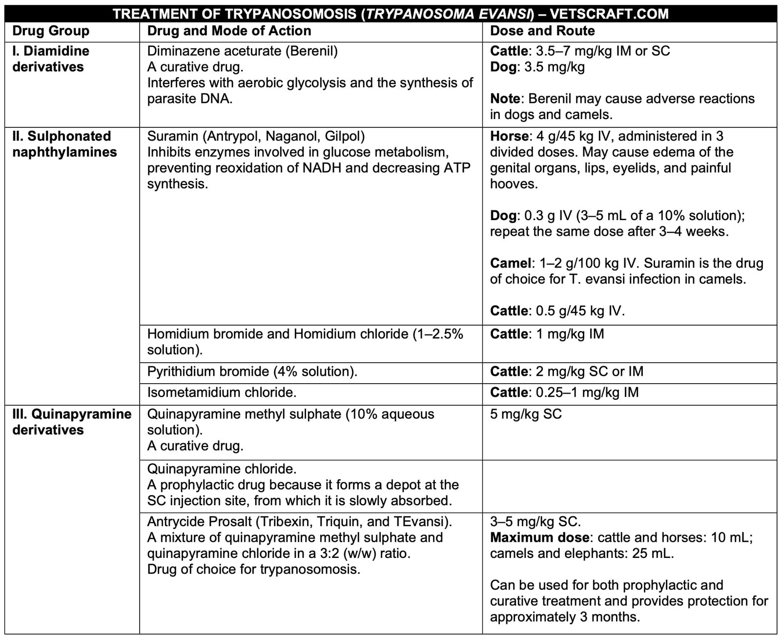
Treatment of Trypanosoma evansi infection is based on the animal species, stage of the disease, and severity of clinical signs. Several trypanocidal drugs are available for both curative and prophylactic use. Early diagnosis and prompt treatment improve the prognosis and help reduce economic losses.
The commonly used drugs, their mode of action, recommended dosage, and routes of administration are summarized below.
Control
- Chemotherapy.
- Vector Control (Removal of vegetation surrounding water sources).
- Destruction of wild reservoir animals.
- Livestock management and quarantine practices.
- Chemoprophylaxis before the onset of the rainy season.
- Rearing trypanotolerant breeds (e.g., N’Dama).

