Trypanosoma: Morphology, Classification, Developmental Stages, Salivaria & Stercoraria
Trypanosoma is a genus of hemoflagellate protozoan parasites belonging to the family Trypanosomatidae. These parasites infect a wide range of domestic animals, wildlife, and humans, causing diseases of significant veterinary and medical importance. Trypanosomes are characterized by their distinctive morphology, multiple developmental stages, and complex transmission through insect vectors or, in the case of T. equiperdum, by venereal transmission.
Trypanosomes are extracellular parasites of the circulatory system and tissue fluids, except Trypanosoma cruzi, which may invade host cells. All species of trypanosomes, except T. equiperdum, are transmitted either mechanically or cyclically by blood-sucking insects. T. equiperdum is transmitted through coitus (venereal transmission). The disease caused by trypanosomes, known as trypanosomosis, is one of the most important diseases of livestock and humans.
Classification of Trypanosoma
- Kingdom: Protozoa
- Phylum: Euglenozoa
- Class: Kinetoplastea
- Order: Trypanosomatida
- Family: Trypanosomatidae
- Genus: Trypanosoma
Morphology of Trypanosomes
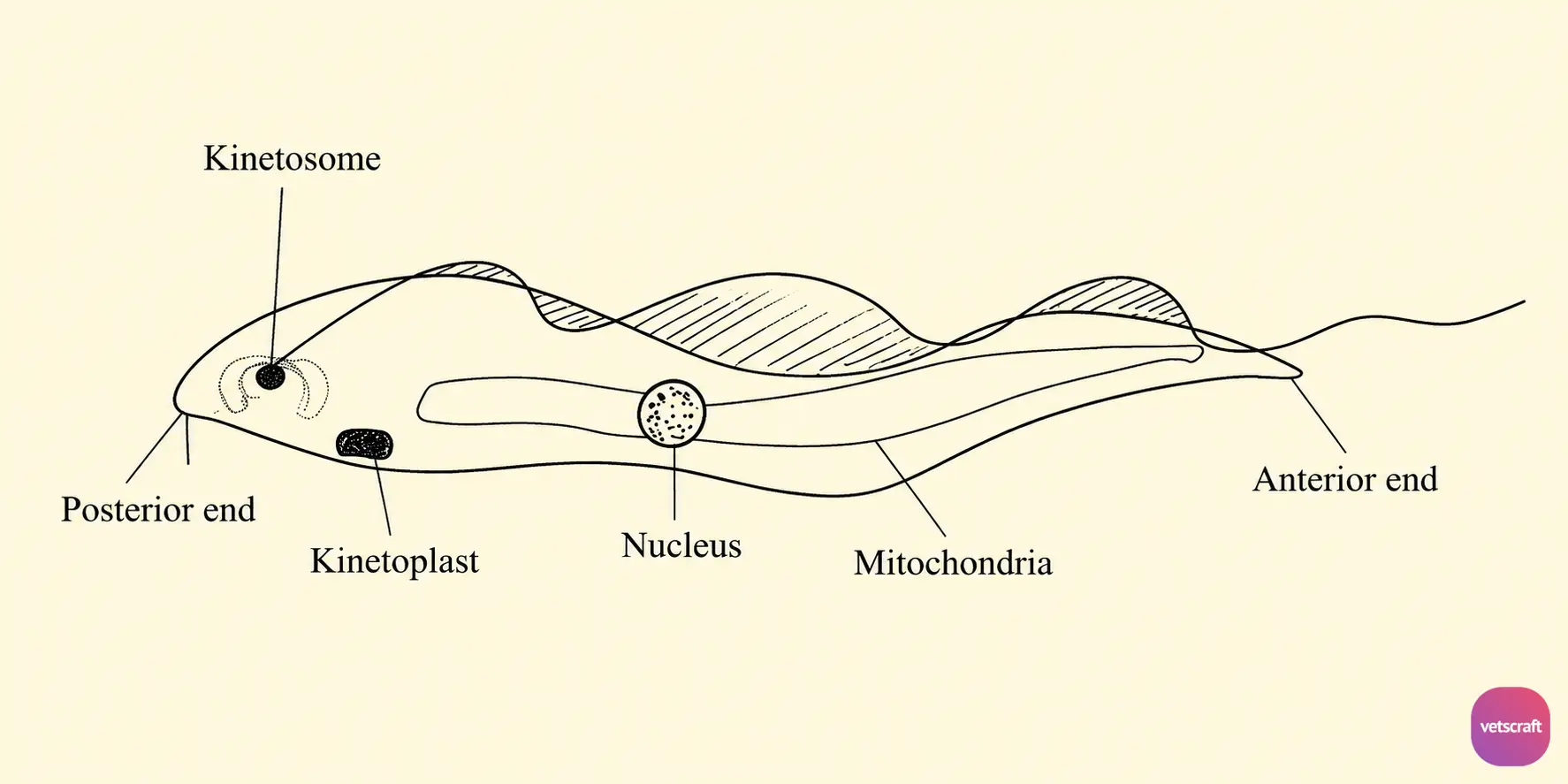
- The body is leaf-shaped with a centrally located vesicular nucleus.
- They have a single flagellum arising from a kinetosome (basal body). In some species, the flagellum passes along the side of the body toward the anterior end and is attached to the body at several points by means of an undulating membrane.
- Posterior to the kinetosome, there is a rod-shaped or spherical structure called the kinetoplast, which contains DNA. In some trypanosomes (Trypanosoma equiperdum), the kinetoplast fails to stain, usually after treatment with acriflavine; these are referred to as dyskinetoplastic or akinetoplastic strains.
- There is a single mitochondrion extending throughout the entire length of the body.
Developmental Stages of Trypanosomes
Trypanosomes are heteroxenous parasites. They pass through different developmental stages in both vertebrate and invertebrate hosts.
The developmental stages of trypanosomes include:
- Amastigote
- Promastigote
- Epimastigote
- Trypomastigote
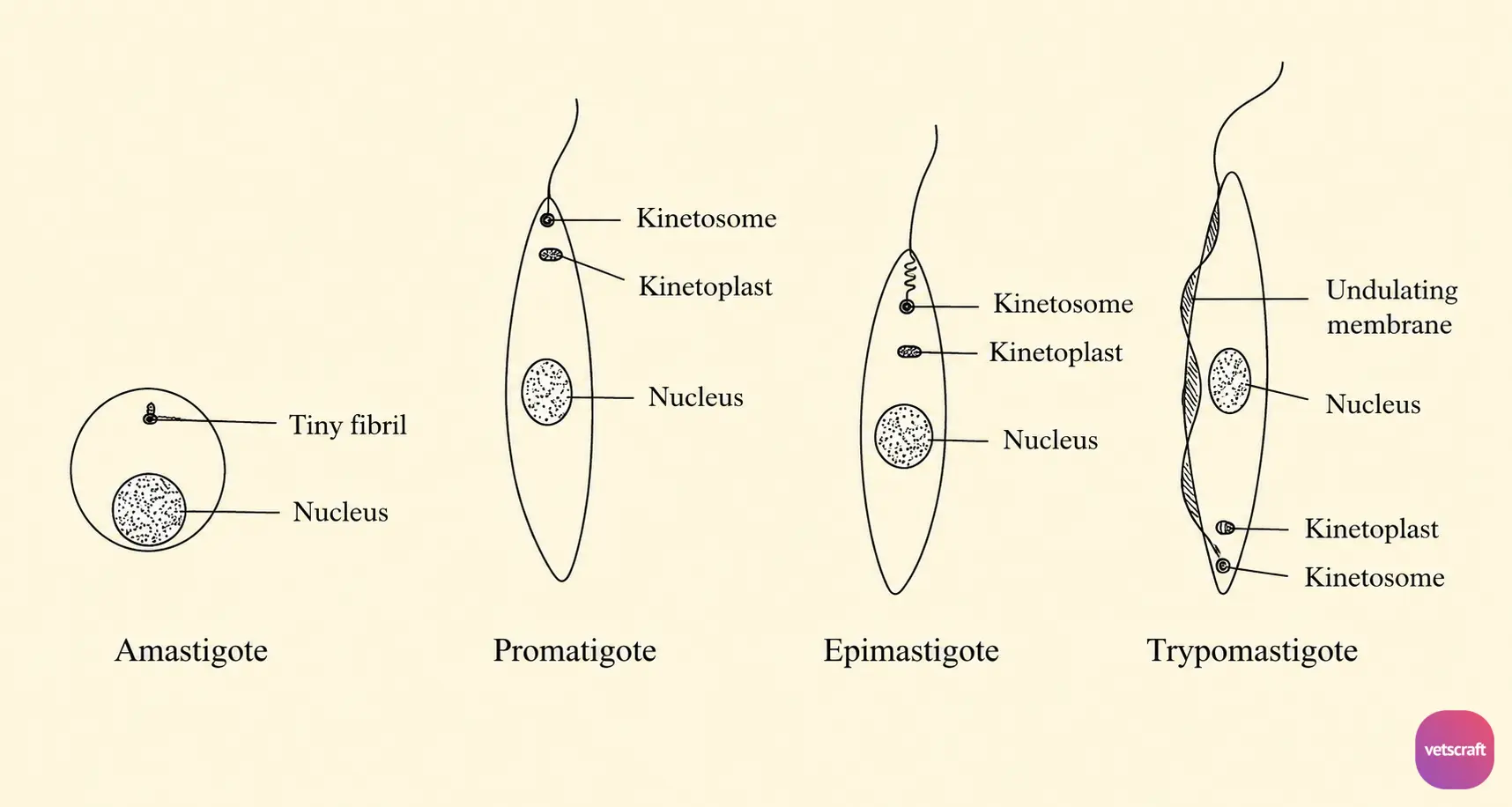
1. Amastigote/Micromastigote (Leishmania Stage)
- The body is rounded.
- The flagellum may be absent; if present, it may be reduced to a tiny fibril.
- The amastigote stage is found in the vertebrate host.
2. Promastigote Stage (Leptomonad Stage)
In this stage, the kinetoplast and kinetosome are located at the tip of the anterior end, and the undulating membrane is absent. This stage is found in the arthropod (invertebrate) host.
3. Epimastigote Stage (Crithidial Stage)
In the epimastigote stage, the kinetoplast and kinetosome lie just anterior to the nucleus, and the undulating membrane extends forward. Although this stage may occur in vertebrates, it is primarily found in arthropods.
4. Trypomastigote Stage
The body is blade-shaped, and the kinetoplast and kinetosome are located near the posterior end. A well-developed undulating membrane is present. This stage is found in the vertebrate host and also occurs in arthropods as the infective stage for the vertebrate host.
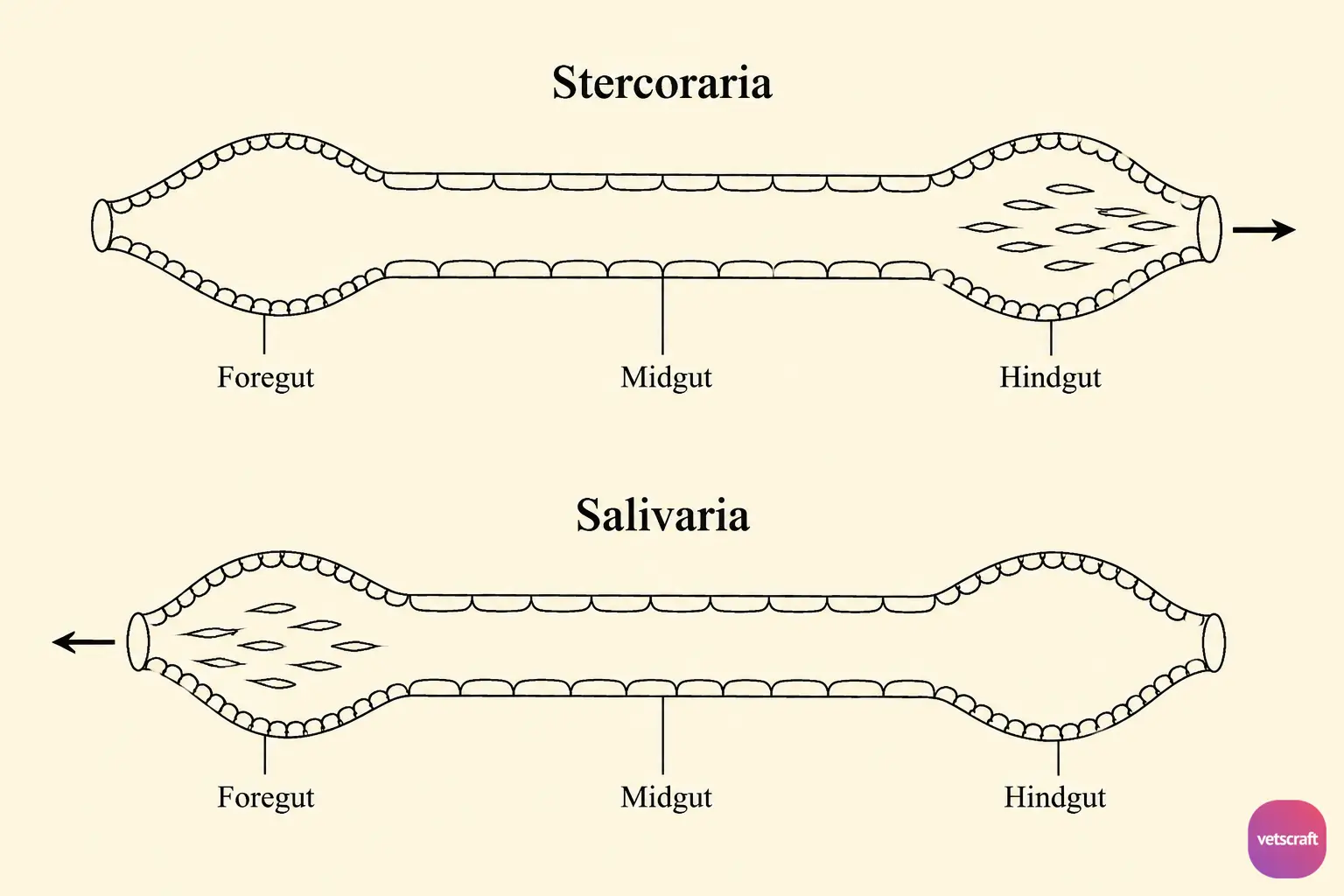
Based on the site where trypanosomes develop within insect vectors, they are classified into Salivaria (anterior station development) and Stercoraria (posterior station development).
Stercoraria
In Stercoraria (posterior station development), the trypanosomes grouped under this category are known as stercorarian trypanosomes. In this group, multiplication occurs in the hindgut of the insect, where the epimastigote stage multiplies and eventually produces large numbers of metacyclic trypomastigotes.
These metacyclic trypomastigotes accumulate in the hindgut of the arthropod and are subsequently passed in the feces. Infection of the definitive host occurs through fecal contamination.
Important characteristics of stercorarian trypanosomes:
- A free flagellum is present in the trypomastigote stage.
- The kinetoplast is large and non-terminal.
- The posterior end of the body is pointed.
- Members of this group are generally less pathogenic.
Salivaria
Salivaria (anterior station development) includes the trypanosomes grouped under this category, which are known as salivarian trypanosomes.
In this group, multiplication of trypanosomes occurs in the midgut (within the peritrophic membrane) of the insect, where the trypomastigote stage multiplies and eventually produces large numbers of metacyclic trypomastigotes.
These metacyclic trypomastigotes accumulate in the mouthparts and salivary glands. Infection of the definitive host occurs through inoculation during the feeding of the arthropod vector.
Important characteristics of Salivaria:
- A free flagellum may be present or absent.
- The kinetoplast is terminal or subterminal.
- The posterior end of the body is usually blunt.
- Members of this group are highly pathogenic.
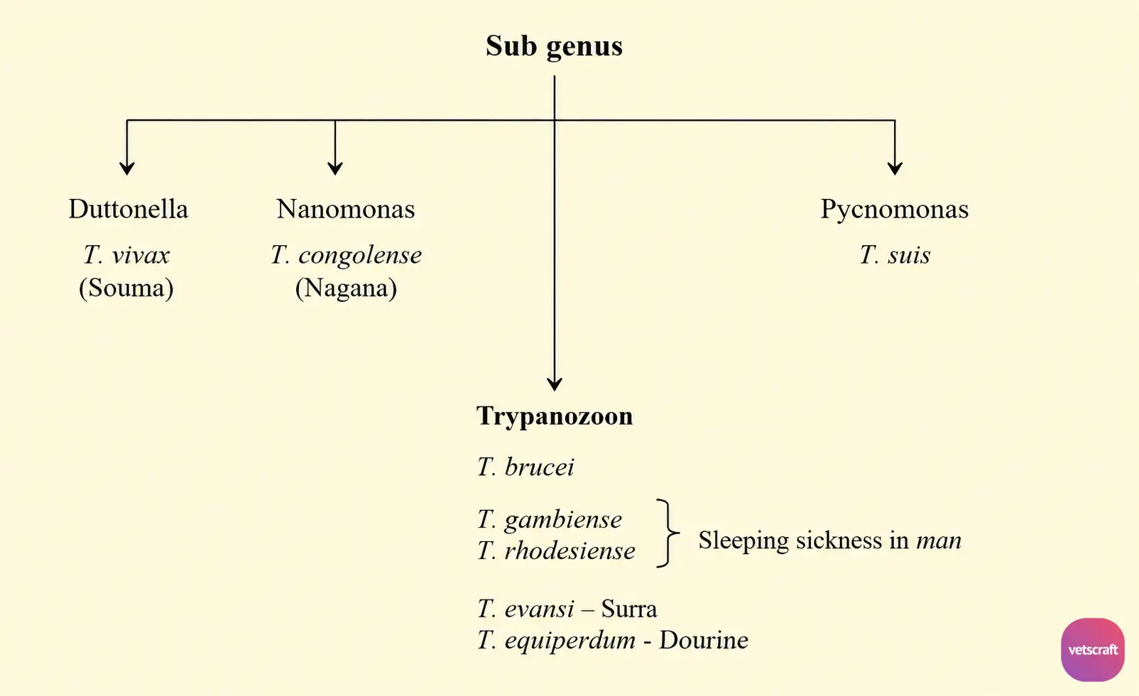
Subgenus of Trypanosoma