
Trypanosoma equiperdum (Dourine in Horses): Morphology, Clinical Signs, Diagnosis & Treatment
Trypanosoma equiperdum is a flagellated hemoprotozoan parasite belonging to the family Trypanosomatidae and is the causative agent of dourine, a chronic venereal disease of equids. Unlike most pathogenic trypanosomes, T. equiperdum is transmitted primarily through natural mating and does not require an arthropod vector for its life cycle.
The disease mainly affects horses and donkeys, causing progressive lesions of the genital tract, skin, and nervous system, which can ultimately result in paralysis and death if left untreated.
Parasite Overview
- Common Name: Dourine parasite
- Disease Caused: Dourine (Equine syphilis)
- Definitive Host: Horses and donkeys
- Intermediate Host: None
- Vector: None (transmitted directly during coitus)
- Distribution: Asia, Africa, parts of Europe, South America, the Middle East, India, and countries of the former USSR
- Predilection Site: Genital mucosa, lymphatic system, blood, and nervous tissue
- Infective Stage: Trypomastigote
- Life Cycle: Direct (monoxenous); no intermediate host or biological vector is involved. Transmission occurs primarily through venereal contact during mating.
- Zoonotic Importance: No; T. equiperdum is not considered zoonotic.
Taxonomical Classification
- Kingdom: Protista
- Phylum: Euglenozoa
- Class: Kinetoplastea
- Order: Trypanosomatida
- Family: Trypanosomatidae
- Genus: Trypanosoma
- Subgenus: Trypanozoon
- Species: Trypanosoma equiperdum
Morphology
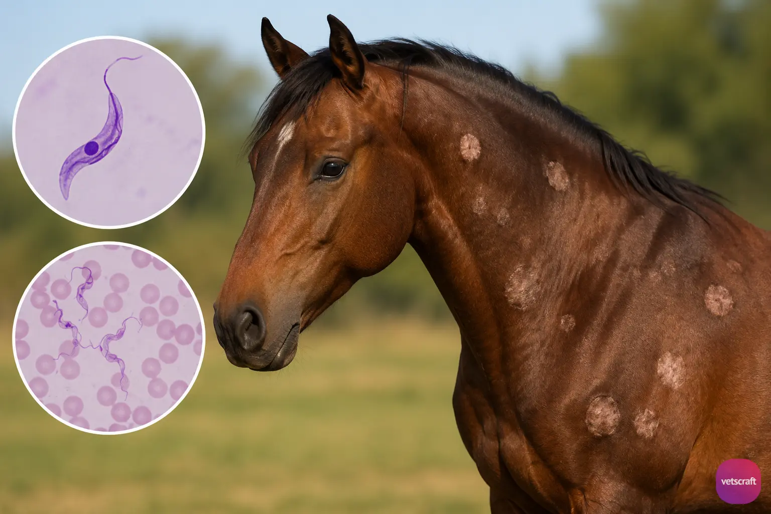
The morphology of Trypanosoma equiperdum is similar to that of Trypanosoma evansi. It occurs as a trypomastigote in the host and possesses an elongated spindle-shaped body, a single flagellum, an undulating membrane, a centrally located nucleus, and a small posterior kinetoplast.
Clinical Manifestations
The clinical manifestations of dourine are divided into three phases:
- Stage of edema
- Urticarial phase
- Phase of paralysis
1. Stage of Edema
The stage of edema begins with a mucoid vaginal or urethral discharge, varying degrees of nymphomania, mild fever, and edema of the genitalia. In stallions, the prepuce and scrotum are swollen, and the edema may extend to the chest.
In mares, the vaginal mucosa is hyperemic, and ulcers may be present. Depigmented areas may occur on the vulva and penis. This phase lasts four to six weeks. In severe cases, frequent micturition and even abortion in pregnant mares may occur.
2. Urticarial Phase
Characteristic urticarial plaques (silver dollar plaques) develop on the neck, chest, abdomen, and croup. These plaques are circular, edematous, and painless and typically persist for a few days before disappearing, only to reappear elsewhere on the body.
3. Phase of Paralysis
The muscles of the face and nostrils are affected first, followed by the hind limbs and the rest of the body. This leads to incoordination, followed by complete paralysis. Dourine is usually fatal unless treated appropriately.
Diagnosis
Demonstration of parasites in smears prepared from the fluid squeezed from urticarial plaques and the mucous membranes of the genitalia.
Serological tests such as the complement fixation test (CFT), indirect fluorescent antibody test (IFAT), and ELISA are also used for the diagnosis of dourine, particularly in chronic infections where parasites are difficult to detect.
Treatment
The treatment of Trypanosoma equiperdum infection is similar to that of Trypanosoma evansi. Trypanocidal drugs such as diminazene aceturate, quinapyramine sulfate, or suramin may be used depending on regional recommendations and availability. In many countries, however, affected animals are culled as part of disease control programs because dourine is a notifiable disease.

