
Tritrichomonas foetus: Morphology, Transmission, Pathogenesis, Diagnosis, Treatment & Control
Tritrichomonas foetus is a flagellated protozoan parasite that primarily infects the reproductive tract of cattle, causing bovine trichomonosis, a sexually transmitted disease of major economic importance in the cattle industry. Infection is characterized by infertility, early embryonic death, repeat breeding, pyometra, and occasional abortion in cows, while infected bulls usually remain asymptomatic lifelong carriers, serving as the primary reservoir of infection. Although artificial insemination has significantly reduced its prevalence in many countries, the disease continues to affect naturally bred herds worldwide.
This article provides a comprehensive overview of its taxonomy, epidemiology, clinical significance, laboratory diagnosis, therapeutic approaches, and preventive measures in cattle.
Parasite Overview
- Host: Cattle. It may also occur in zebus, pigs, horses, and deer, but pathogenic effects are observed only in cattle.
- Predilection Site: Reproductive system
- Disease: Venereal disease, bovine trichomonosis
- Distribution: Worldwide. It was once considered an economically important disease in dairy herds, but its incidence has declined significantly due to the widespread use of artificial insemination.
Taxonomical Classification
- Kingdom: Protista
- Phylum: Metamonada
- Class: Parabasalia
- Order: Trichomonadida
- Family: Tritrichomonadidae
- Genus: Tritrichomonas
- Species: Tritrichomonas foetus
- Common Name: Bovine trichomonad, feline trichomonad
Morphology
The organism is roughly pear-shaped, measuring 10–25 µm long and 3–15 µm wide. It has a rounded anterior end and a somewhat pointed posterior end.
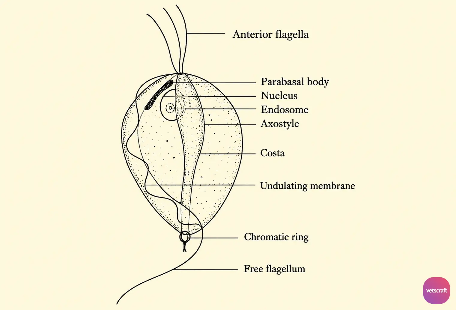
There is a single nucleus located at the anterior end and a sausage-shaped parabasal body. Three anterior flagella are present, and a posterior flagellum extends along the undulating membrane and trails behind as a free flagellum.
The axostyle is well developed and emerges from the body through a chromatic ring. It has a prominent costa but no pelta.
Trichomonads multiply by longitudinal binary fission. There is no sexual reproduction or encystment stage in the life cycle.
Based on the pathogenicity of Tritrichomonas, it has been differentiated serologically into three distinct strains: Belfast (mild), Manley (intermediate), and Brisbane (pathogenic).
Epidemiology
Bovine trichomonosis is a venereal disease transmitted by coitus. T. foetus may or may not survive in frozen semen, depending on the storage conditions. Rapid freezing and high salt concentrations are detrimental to the organism.
Once a bull becomes infected, it carries the infection for life and serves as a permanent source of infection. In females, however, the infection is self-limiting, and the parasite gradually disappears, allowing spontaneous recovery. Following recovery, cows usually conceive and undergo normal gestation.
Transmission
Under natural conditions, the infection is transmitted during coitus. It may also be transmitted through artificial insemination and gynecological examination of cows.
Pathogenesis
In bulls, the most common site of infection is the preputial cavity. Early clinical signs include pain during urination and reluctance to breed.
A mucopurulent discharge may be present. These signs may disappear within one to two weeks after infection, but the bull remains infected for life. Therefore, infected bulls are regarded as permanent sources of infection.
In females, the organisms are introduced into the vagina during coitus, where they multiply and cause vaginitis. The maximum number of organisms is present in the vagina 14–18 days after infection, after which they invade the uterus through the cervix.
Invasion of the uterus leads to placentitis, detachment of the placental membranes, and death of the fetus. This results in early abortion, usually between 8 and 16 weeks of gestation, which is characteristic of bovine trichomonosis.
Because the fetus is very small during early pregnancy, early abortion may go unnoticed. The producer may believe that the animal failed to conceive or has an irregular estrous cycle.
Despite the infection, some animals may conceive normally, carry the calf to full term, and deliver a healthy calf. If the placenta and fetal membranes are completely expelled after abortion, the cow usually recovers spontaneously.
Sometimes the animal does not abort, but the fetus dies and becomes macerated in the uterus, leading to chronic catarrhal and purulent endometritis. As a result, permanent sterility may occur. Affected animals exhibit anestrus and persistent uterine discharge, which may be noticeable when the animal lies down.
Sometimes the cervix remains closed, and in the presence of a persistent corpus luteum, closed pyometra may develop. In such cases, grayish-white, odorless fluid accumulates in the uterus and contains large numbers of trichomonads. These conditions result in anestrus, and the owner may mistakenly assume that the animal is pregnant.
Immunity
Bulls that recover from infection become immune to reinfection. However, they usually harbor the organisms for life and remain a continuous source of infection for cows. In cows, non-pregnant animals tend to recover from infection without treatment.
In T. foetus infections, the following types of antibodies are produced:
- Circulating Humoral Antibody: Produced against antigens that reach the systemic circulation from the uterus. In natural infections, this antibody is produced only when large numbers of trichomonads are present in the uterus.
- Uterine Antibody: Induced by trichomonads in the uterus. It is responsible for the elimination of organisms from the uterus in mild infections.
- Vaginal Antibody: Produced by the vaginal mucosa when large numbers of organisms are present in the vagina.
These antibodies are most readily detected using an agglutination technique.
Diagnosis
- History of early abortion.
- High incidence of cows returning to estrus (repeat breeders). Animals fail to conceive even after several breeding attempts.
- Definitive diagnosis is based on the demonstration of organisms in vaginal or uterine discharge or in the stomach contents of an aborted fetus.
Collection of Samples
- In abortion cases, stomach contents of the aborted fetus can be collected.
- Samples can be obtained by washing the vagina with physiological saline in females. In bulls, preputial washings are the best source of material.
- During sample collection, avoid contamination with fecal material, as this may introduce intestinal protozoa into the sample and cause confusion with T. foetus.
Examination of Samples
Organisms may be observed directly by examining purulent mucus in heavy infections. In mild infections, the sample may be allowed to sediment or be centrifuged before examination.
The sample should be kept warm throughout processing, and warm saline should be added before examination.
Immunological Test
The mucus agglutination test is the most satisfactory immunodiagnostic test. Mucus samples should be collected from the vagina of the cow, preferably a few days after estrus, using a sterile glass tube 50 cm in length and 9 mm in diameter, bent at an angle of 150° approximately 9 cm from one end.
The mucus is mixed with glucose saline and serially diluted. The test antigen should contain T. foetus (produced by in vitro culture) at a concentration of 100,000 organisms/mL.
Cultivation of Organism
Two media are commonly used for the cultivation of trichomonads: diphasic glucose broth-serum medium and Diamond medium, which contains trypticase, yeast extract, maltose, cysteine, and serum.
Treatment of Bulls
Treatment of bulls is difficult because it is tedious, time-consuming, and must be repeated several times. Therefore, culling is generally recommended. However, if the bull has high genetic or economic value, treatment may be attempted.
Treatment Procedure
- First, the retractor penis muscle is relaxed by pudendal anesthesia to expose all parts of the preputial cavity to the drug.
- The penis is then washed with a dilute detergent solution and dried.
- After drying, 1% acriflavine ointment or solution is introduced into the preputial cavity and massaged for 15–20 minutes.
- A 1% Berenil solution (100–150 mL) is injected into the prepuce and retained for 15 minutes while the penis and prepuce are thoroughly massaged.
- Dimetridazole: 50 mg/kg orally once daily for 5 days.
- Dimetridazole: 50 mg/kg as a single intravenous injection.
Control
- Artificial insemination should be practiced for breeding.
- Avoid the use of communal bulls for breeding.
- Infected bulls should be culled.
- Breeding rest should be provided to affected cows.

