
Coccidiosis in Dogs and Cats (Isospora): Transmission, Clinical Signs, Diagnosis, Treatment and Prevention
Coccidiosis in dogs and cats is a protozoal intestinal disease caused primarily by Isospora species (also known as Cystoisospora in the current taxonomy). Although infections are often subclinical in healthy animals, young, stressed, or immunocompromised dogs and cats may develop diarrhea and other gastrointestinal signs. The disease is transmitted through ingestion of sporulated oocysts or infected transport hosts and is an important cause of enteric disease in young animals.
Two species, Isospora canis and Isospora felis, are commonly found in dogs and cats, respectively. Wild animals such as wild cats, tigers, and lions may also be affected.
In addition, other animals such as mice, rats, golden hamsters, calves, cattle, and dogs may become infected and act as transport hosts, in which hypnozoites (bradyzoite cysts) develop in the lymph nodes, liver, spleen, and occasionally in the skeletal muscles.
Predilection Site: Small intestine, cecum, and occasionally the colon of dogs and cats.
Structure of the Oocyst
- Ovoid, 32–53 × 26–43 µm.
- The oocyst wall is smooth, yellowish to pale brown, single-layered, and apparently lined by a thin membrane.
- No micropyle.
- No residuum or polar granule.
- Sporocysts are ellipsoidal without a Stieda body.
- Sporozoites are sausage-shaped.
- Sporulation Time: 2 days or less.
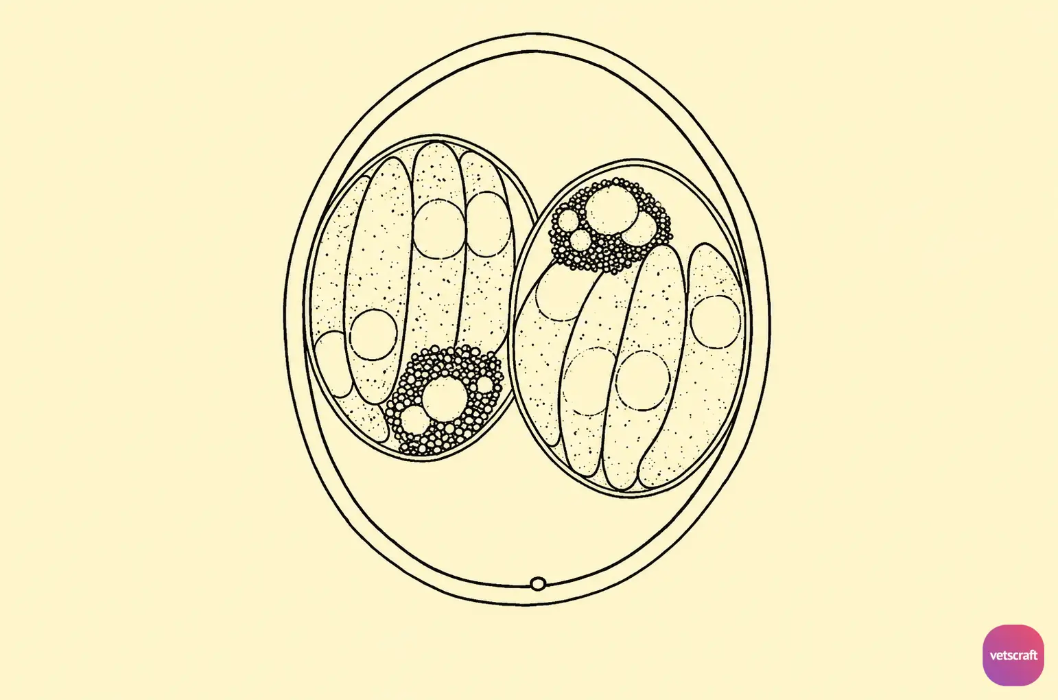
The sporulated oocysts of Isospora contain two sporocysts, each containing four sporozoites.
Transmission
Dogs and cats become infected by ingesting sporulated oocysts or tissue cysts in transport hosts. Following ingestion, three generations of schizogony and gametogony take place in the intestinal wall.
The first-generation schizont produces 16–17 banana-shaped merozoites. Each merozoite, in turn, produces 2–10 merozoites, forming the second-generation schizont. Third-generation schizonts are formed on the sixth day of infection. Sexual development (gametogony) occurs mainly in the small intestine 7–8 days after infection but may also occur in the large intestine. The prepatent period is 7–8 days.
Pathogenicity and Clinical Signs
Isospora spp. are normally not pathogenic; however, if animals are under stress, immunosuppression, or corticosteroid (prednisolone or dexamethasone) therapy, they may develop clinical disease. Affected animals may show diarrhea, anorexia, emaciation, and death in severe cases. Pathological changes consist of catarrhal enteritis in mild cases and hemorrhagic enteritis in heavy infections.
Diagnosis
- Clinical signs.
- Fecal examination.
- Examination of intestinal scrapings shows the presence of a large number of developmental stages.
This condition should be differentiated from ancylostomosis and canine parvovirus infection.
Treatment
- Nitrofurazone: 1.4 mg/kg three times daily for 10 days.
- Mepacrine at 0.01 g/kg is effective in cats.
- Sulfamethazine is effective in dogs at 0.066 g/kg.
- Secondary bacterial infections can be treated with tetracycline compounds.
Prevention
- Maintain proper kennel hygiene.
- Clean kennels with boiling water.
- Isolate and treat affected animals.

