
Demodex canis in Dogs: Morphology, Life Cycle, Clinical Signs, Diagnosis & Treatment
Demodex canis is a host-specific mite that normally inhabits the hair follicles and sebaceous glands of dogs. It belongs to the Order Trombidiformes (Prostigmata) and Family Demodecidae. Under certain conditions, excessive proliferation of these mites can result in canine demodicosis, which may occur in localized or generalized forms. This post covers the morphology, life cycle, transmission, predisposing factors, clinical forms, diagnosis, and treatment of Demodex canis infestation in dogs.
- Location: Species of this genus are highly host-specific and live as commensals. They are found in the hair follicles, sebaceous glands, and meibomian glands of dogs.
- Disease: Red mange, follicular mange, demodectic mange, canine demodicosis
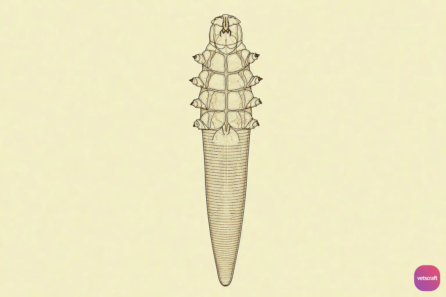
Morphology
- Burrowing-type mites; they are minute mites with an elongated, carrot-shaped body.
- They have a head, a thorax that bears four pairs of short and stumpy legs, and an elongated abdomen, which is transversely striated on the dorsal and ventral surfaces.
- The abdomen is at least half the length of the body.
- The penis protrudes from the dorsal surface in males, and the vulva is on the ventral surface in females.
- Eggs are spindle-shaped.
Life Cycle
- The entire life cycle is spent on the host and consists of the egg, larva, protonymph, deutonymph, and adult stages.
- The life cycle is completed in 18–24 days in the hair follicles or sebaceous glands in a characteristic head-downward posture.
- Males occur at or near the skin surface, whereas fertilized females oviposit 20–24 eggs in the hair follicles.
- Larvae and nymphs are swept by the sebaceous flow to the mouth of the follicle, where they mature and repeat the cycle.
- The species are unable to survive off their host.
Mode of Transmission
Infection is transmitted by direct contact or experimentally by the application of infected skin.
Neonatal contact with an infected mother during suckling establishes the infection within the first few days of life, usually within 72 hours after birth. Most infections are acquired during the early weeks of life.
A hereditary predisposition to generalized demodicosis has been demonstrated in some dogs, although the underlying immunologic mechanisms are not fully understood. Littermates may have an increased risk of developing generalized demodicosis because of their shared genetic predisposition.
Predisposing Factors
A number of factors predispose dogs to clinical disease:
- Dogs aged 3–9 months are mostly affected, but adults in poor condition or with inadequate nutrition may also be affected.
- Intercurrent infections, especially viral infections.
- Excessive use of alkaline soaps or shampoos.
- Genetic predisposition, hyperadrenocorticism, stress, endoparasitic infections, debilitating diseases (neoplasia), and corticosteroid treatment (immunosuppressive).
Breed Predisposition
Short-haired dogs (White Terrier, Bull Terrier, Boxer, Dobermann) and long-haired dogs (German Shepherd, Afghan Hound, Collie) are susceptible to infection.
Pathogenesis
Impaired cell-mediated immunity and other immunologic abnormalities may predispose dogs to excessive proliferation of Demodex mites. The precise immunologic mechanisms underlying generalized demodicosis are not fully understood.
A hereditary predisposition to generalized demodicosis has been demonstrated in some dogs, although the underlying immunologic mechanisms are not fully understood.
Demodectic mange may flare up when dogs are given immunosuppressive therapy for other conditions.
Clinical Features of Canine Demodicosis
Demodectic mange has been classified based on clinical condition as:
- Juvenile-onset demodicosis: It occurs in young dogs and may present as localized or generalized disease. Localized lesions are often self-limiting, whereas generalized disease requires treatment. Immunosuppressive therapy may exacerbate the disease.
- Adult-onset Demodicosis: It is often associated with an underlying disease or immunosuppressive condition and may be accompanied by secondary bacterial pyoderma. Clinical signs may include alopecia, erythema, scaling, crusting, and pustules. The skin often becomes hyperpigmented in chronic cases. It typically occurs in adult dogs, usually older than 5 years of age.
- Localized Form: It is found on the eyelids, periorbital fossa, nose, muzzle, forelimbs, and ears.
- Generalized Form: It occurs over all parts of the body.
- Pododemodicosis: It involves the paws and may occur as a localized or generalized form of demodicosis. It may be accompanied by secondary bacterial infection and inflammation.
Squamous Form
The squamous form is a less serious form and causes a dry reaction. The hair follicles are distended with mites and cellular debris, and the follicular epithelium is atrophic.
Hyperkeratosis progressively develops, and pieces of cornified material exfoliate from the surface. The hair may become separated and shed, splintered, or disintegrated. Changes in the sebaceous glands vary; they may be atrophied or hypertrophied.
Hyperpigmentation occurs, giving the skin a coppery-red color. The condition progresses until large areas of the body are affected. Animals may show alopecia, thickened and wrinkled skin, and a “mousy” odor.
Pustular Form
The pustular form is usually preceded by the squamous form. It is a serious form that results from bacterial invasion (Staphylococcus sp.) of the dermis.
Extensive dermal infiltration of polymorphonuclear leukocytes, lymphocytes, and plasma cells is seen.
Pustule or abscess formation with marked inflammation occurs. Serum, pus, and blood ooze from the lesions, thereby giving rise to the common name “red mange.” In extensive forms of canine demodicosis, death results from toxemia or emaciation.
Pruritus may be absent or mild in uncomplicated demodicosis but can become marked when secondary bacterial infection or other concurrent skin disease is present.
Diagnosis
- By deep skin scrapings of the contents of pustules or abscesses.
- Scrapings must be deep enough to ensure capillary oozing and sampling of the hair follicles.
- The skin may be softened with a weak solution of KOH/NaOH to assist with scraping.
Treatment
- Isoxazolines are currently the preferred treatment for generalized canine demodicosis. Effective options include afoxolaner, fluralaner, lotilaner, and sarolaner, administered at their labeled flea/tick-control dose and interval.
- Moxidectin can be used as a topical spot-on, particularly in combination with imidacloprid. It is an established alternative treatment for canine demodicosis.
- Milbemycin oxime can be administered orally at 1–2 mg/kg once daily for generalized demodicosis.
- Ivermectin can be administered orally at 0.3–0.6 mg/kg once daily as an alternative treatment for generalized demodicosis.
- Doramectin has also been used for canine demodicosis, with published protocols including 0.6 mg/kg subcutaneously once weekly.
- Amitraz may be used as a topical dip for generalized demodicosis where an appropriate veterinary product is available; historical protocols use concentrations around 0.025–0.06% at weekly or biweekly intervals.
- Secondary bacterial pyoderma should be treated concurrently with appropriate topical and/or systemic antimicrobial therapy.
- Treatment should be continued until clinical resolution and repeated skin examinations demonstrate parasitological cure, with monthly deep skin scrapings commonly used to monitor response. Underlying diseases associated with adult-onset demodicosis should be investigated and treated to improve the response to acaricidal therapy.
- Corticosteroids should never be given in demodicosis.

