
Otodectes cynotis (Ear Mites) in Dogs and Cats: Morphology, Life Cycle, Diagnosis & Treatment
Otodectes cynotis, commonly known as the ear mite, is a highly contagious surface-dwelling mite that primarily infests the external ear canals of cats and dogs. It is particularly common in cats and can cause otitis externa characterized by intense pruritus, head shaking, erythema, and accumulation of dark, waxy to crusty ceruminous debris. The mite belongs to the Order Sarcoptiformes, Suborder Astigmata, and Family Psoroptidae.
Otodectes cynotis does not burrow into the skin. Instead, the mites live on the surface of the skin within the external ear canal, where they feed on epithelial debris, tissue fluids, and exudate. Although the ears are the primary site of infestation, mites may occasionally be found on other areas of the body, including the head, neck, dorsum, tail, and limbs.
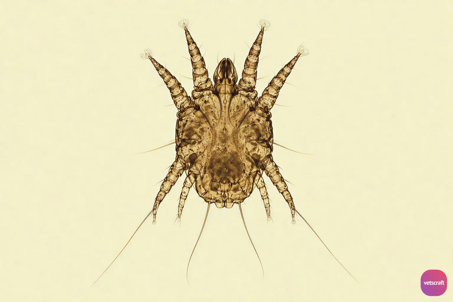
- Common name: Ear mite, ear canker mite
- Host: Primarily cats and dogs; also reported in ferrets, foxes, and other carnivores
- Site of infestation: External ear canal, occasionally other areas of the skin
- Disease: Otodectic mange, otoacariasis, parasitic otitis externa
Morphology
Otodectes cynotis is a relatively large mite compared with many other mites affecting dogs and cats. Adult mites are approximately 300–500 µm in length and may sometimes be visible without magnification as small, whitish, mobile organisms.
- The body is oval to roughly rounded and is divided into the anterior gnathosoma and posterior idiosoma.
- Adult mites possess four pairs of legs.
- The legs terminate in short, unsegmented pedicels bearing suckers (caruncles).
- In females, suckers are present on the first and second pairs of legs, while the fourth pair of legs is markedly reduced.
- Males have suckers associated with all four pairs of legs and possess characteristic copulatory structures on the ventral surface.
- The gnathosoma is relatively short and conical compared with the elongated gnathosoma of Demodex species.
- The characteristic short pedicels and terminal suckers are important morphological features for differentiating Otodectes from some other psoroptid mites.
Hosts and Distribution
Otodectes cynotis has a worldwide distribution. It primarily affects domestic cats and dogs but has also been reported in other carnivorous hosts, including ferrets and wild canids and felids.
Infestation is particularly common in cats, especially young and free-roaming cats. However, dogs can also develop clinically significant otoacariasis. The mite can spread rapidly among animals living in close contact.
Life Cycle
The entire life cycle of Otodectes cynotis occurs on the host and is completed primarily within the external ear canal. The complete life cycle generally takes approximately 3 weeks under favorable conditions.
- The developmental stages include egg, larva, protonymph, deutonymph, and adult.
- Adult females deposit eggs that become attached to the epidermal surface of the ear canal.
- The egg develops into a six-legged larva.
- The larva molts into an eight-legged protonymph.
- The protonymph subsequently develops into a deutonymph.
- The deutonymph molts into the adult stage.
- Adults reproduce within the ear canal, and the cycle continues.
Mode of Transmission
Transmission occurs primarily through direct contact with an infested animal. Close contact between cats and dogs allows mites to move from one host to another.
- Direct contact with an infested animal is the principal route of transmission.
- Transmission may occur readily between littermates and between a mother and her offspring.
- Contaminated bedding, grooming equipment, and other fomites may contribute to transmission, although off-host survival is generally limited.
- In multi-animal households, untreated contact animals can serve as a source of reinfestation.
Pathogenesis
Otodectes cynotis is a surface-dwelling mite and does not burrow into the skin. The mites feed on epithelial debris, tissue fluid, and exudate present within the external ear canal.
The mechanical irritation caused by mite activity, together with the host’s hypersensitivity response to mite antigens, results in inflammation of the ear canal. The degree of clinical disease does not always correlate directly with the number of mites present.
Persistent scratching and head shaking can cause self-trauma to the pinna and surrounding skin. In severe cases, repeated trauma may contribute to excoriation, alopecia, ulceration, and development of an aural hematoma.
The inflammatory response and accumulation of cerumen and debris can create conditions favorable for secondary bacterial or yeast overgrowth, resulting in concurrent otitis externa.
Clinical Signs
Clinical signs vary considerably among individual animals. Some animals may harbor mites with minimal or no obvious clinical signs, whereas others develop marked pruritus and severe otitis externa.
- Intense pruritus of the ears
- Frequent scratching of the ears
- Head shaking
- Rubbing the ears against surfaces
- Erythema of the external ear canal
- Accumulation of dark brown to black, waxy or crusty ceruminous debris
- Serous or purulent discharge when secondary infection is present
- Excoriations and alopecia of the pinnae due to self-trauma
- Aural hematoma in severely affected animals
Diagnosis
- Otoscopic Examination: Live mites may sometimes be observed moving within the external ear canal during otoscopic examination. They appear as small, pale, rapidly moving organisms on the surface of the cerumen and skin.
- Microscopic Examination: Microscopic examination of material collected from the external ear canal is a simple and highly useful method of confirming infestation. Ear debris can be collected with a cotton-tipped applicator and examined for mites after transferring the material to a microscope slide with mineral oil.
Treatment
- Acaricidal therapy: Effective acaricides such as selamectin, moxidectin, milbemycin oxime, ivermectin, and appropriate isoxazoline products (fluralaner, sarolaner, etc.).
- Topical therapy: Topical products containing selamectin or other approved acaricidal agents are effective against O. cynotis.
- Ear cleaning: The external ear canal should be appropriately cleaned to remove excessive cerumen, exudate, cellular debris, and dead mites before or during treatment, when clinically indicated.
- Secondary infection: Bacterial or yeast otitis externa should be identified by otoscopic examination and cytology and treated with appropriate topical or systemic therapy when indicated.
- Anti-inflammatory therapy: Animals with marked inflammation or pruritus may require appropriate anti-inflammatory or symptomatic treatment based on the severity of otitis and the presence of secondary infection.

