
Sarcoptes scabiei: Morphology, Life Cycle, Pathogenesis, Clinical Signs, Diagnosis & Treatment
Sarcoptes scabiei is a mite belonging to the family Sarcoptidae that causes scabies in humans and sarcoptic mange in sheep, goats, cattle, pigs, horses, dogs, foxes, rabbits, and other animals. This burrowing mite primarily affects hairless regions or areas with short hair, commonly the face and ears, where it penetrates the skin and causes characteristic lesions and intense pruritus.
The condition is highly contagious and can significantly affect the health and productivity of affected animals.
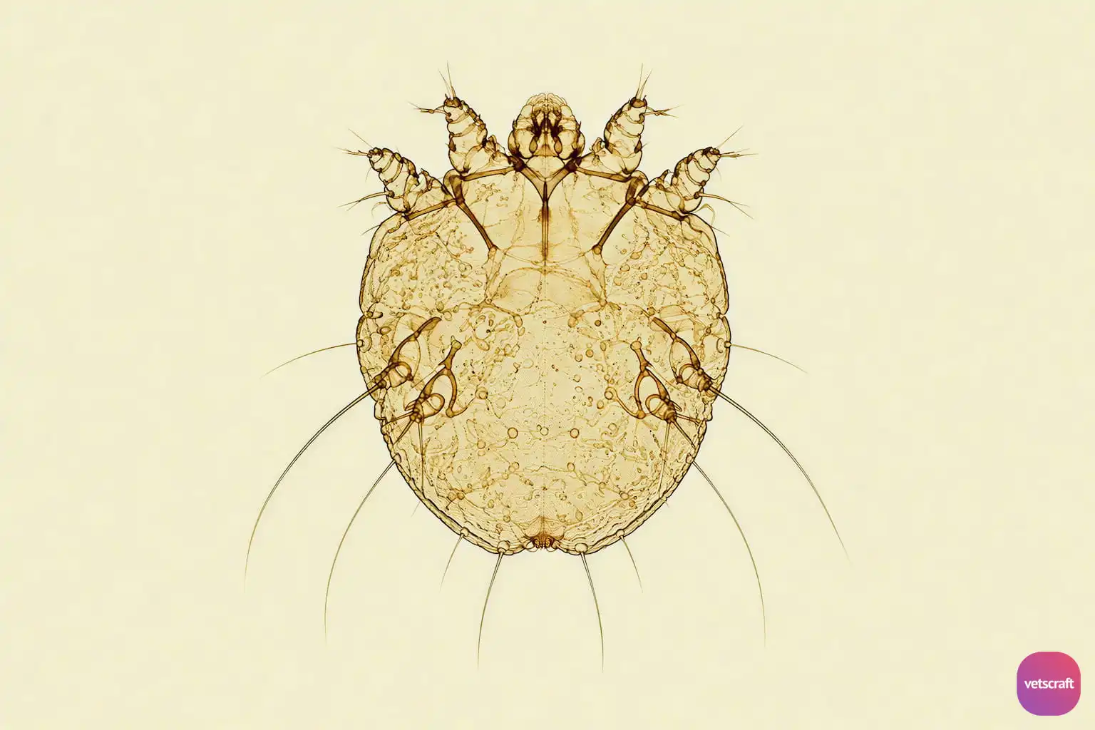
Morphology
- Small parasite with a globose or round shape. It is a burrowing mite.
- The dorsal surface is covered with fine folds, transverse grooves, and small triangular scales.
- The female bears, on either side of the dorsal midline anteriorly, three short spines and, posteriorly, six larger spines with bifid tips.
- Two vertical setae occur on the dorsum of the propodosoma.
- The pedicels are not segmented and bear suckers. The anal opening is terminal.
- All legs in both sexes are short. The third and fourth pairs of legs do not project beyond the body margin and remain within the circumference of the body.
- The tarsi of the first and second pairs of legs in both sexes and the fourth pair in males end in long-stalked, unjointed suckers, while the remaining legs terminate in bristles.
- On the ventral surface, the epimeres (chitinous extensions of the coxae of the legs) are distinct. Those of the first pair of legs are fused into a single rod, while those of the third and fourth pairs of legs are fused to form a lateral bar.
Life Cycle
Females burrow into the skin and lay 40–50 eggs in the tunnels they form. The eggs hatch in 3–5 days into hexapod larvae.
Some larvae escape from the breeding tunnel and wander on the skin, while others remain in the breeding tunnels and develop into nymphs. Those that reach the surface may die, or they may make shallow pockets in the skin tissue, feed, and molt through several nymphal stages. The nymphs can also wander on the surface and make new pockets or extend the molting tunnels. Adult males and females emerge approximately 17 days after the eggs are first laid.
The female remains in her molting pocket until fertilized by a male, then extends it into a breeding tunnel or returns to the skin surface to create a new tunnel and begin laying eggs.
Mature females do not live much longer than a month. Wandering larvae, nymphs, and fertilized females spread the infection on the host and to other hosts. They cannot survive off the host for more than a few days.
The majority remain in the parent tunnel or side pockets, feed voraciously, and molt to become protonymphs and deutonymphs. The nymphs have four pairs of legs and lack a genital opening.
Infection is spread mainly by contact with wandering larvae, nymphs, and fertilized young females.
Transmission occurs through the transfer of impregnated females during person-to-person, skin-to-skin contact. Occasionally, transmission may occur via fomites (e.g., bedding or clothing). Human scabies mites are often found between the fingers and on the wrists.
Pathogenesis
These mites prefer areas with sparse hair, such as the face and ears. In long-standing infections, the mites can spread to all parts of the body.
They pierce the skin to suck lymph fluid and also feed on epidermal scales and skin cells. They cause considerable irritation, itching, and scratching, which worsens the condition.
Crusts form on the skin, which becomes thickened and wrinkled, and the hair falls out. Skin lesions begin to develop within a few days after infection, but the intense itching typical of sarcoptic mite infection does not begin until approximately a month later. The fecal pellets of the mite are responsible for the host inflammatory response and produce marked irritation, which causes intense itching and scratching.
Inflammation of the skin is accompanied by exudates, which coagulate and form crusts on the surface. It is further characterized by excessive keratinization and proliferation of connective tissue. Hair loss becomes widespread.
Sensitization of the host to the mite and its products plays an important role in pathogenesis, with cytokine production and an IgE-mediated hypersensitivity reaction.
Usually, there is no pruritus for 1–3 weeks, after which pruritus appears with an urticarial response.
Animals with severe urticarial reactions recover more quickly than those with less severe reactions. Scraped material from the wrinkled surface will contain myriads of mites.
Clinical Signs
- Dogs: Affects the ears, muzzle, face, and elbows; in severe infestations, it extends over the whole body. It begins as erythema with papule formation, followed by scale and crust formation and alopecia. Intense pruritus leads to self-inflicted trauma and subsequent secondary bacterial infection (pyoderma). After primary infection, dogs may begin scratching before lesions are visible. If lesions are neglected for several months, dogs become progressively weak and emaciated. A foul-smelling odor may occur in severe cases, particularly with secondary infection.
- Cats: Rare in cats and similar to Notoedres. Progressive hair loss occurs on the ears, face, and neck and extends to the abdomen.
- Cattle: Potentially the most severe form of cattle mange. Rare in cattle. The skin becomes scaly and thickened, with marked hair loss. Crusts form on less-haired areas, accompanied by intense pruritus. There will be decreased milk and meat production and reduced skin and hide value.
- Sheep: It preferentially affects the face, ears, axillae, and groin and spreads slowly. Initially, there is erythema, scaling, and intense pruritus. Sheep scratch and rub the head, body, and legs against trees, posts, and walls. Progressive emaciation may occur.
- Goats: Chronic skin disease characterized by irritation, encrustation, hair loss, and excoriation from rubbing and scratching. In long-standing cases, the skin becomes thickened, with nodules developing in less-haired areas such as the muzzle, around the eyes, and inside the ears.
- Horses: Rare. Infection starts on the head, neck, shoulders, and saddle areas. Papule formation and pruritus occur. Thick crusts, scaling, scabs, and hair loss develop, and the infection spreads over the entire body surface. Marked thickening of the skin may occur, with cracking and secondary bacterial infection. In advanced cases, emaciation, cachexia, and eventually death may occur.
- Pigs: The ear is the primary focus, with later spread to the back, flanks, and abdomen. It spreads from carrier sows to suckling piglets and during mating. It is common in piglets 1–3 weeks old. Scratching, loss of condition, small red papules or wheals, and generalized erythema around the eyes and snout occur, resulting in excoriation of the affected areas and formation of brownish scabs. The skin then becomes wrinkled and covered with crusty lesions and becomes thickened. Iron deficiency can exacerbate the condition.
- Rabbits: Lesions first appear on the head and ears and then become generalized. Intense pruritus and hair loss occur. Infection may become severe.
- Humans: Intense pruritus, especially at night, with a papular rash and characteristic burrows. Lesions commonly occur between the fingers, on the wrists, elbows, axillae, waist, buttocks, and genital region. Secondary bacterial infection may develop due to scratching.
Features
- The edges of the ears are often affected first, and rubbing the area readily elicits a scratch reflex.
- There is intense itching, and the condition is highly contagious.
Diagnosis
- Based on clinical signs (intense itching).
- Demonstration of the parasite and/or its eggs in deep skin scrapings; in pigs, mites may also be demonstrated in ear wax.
- This material should be examined in water, glycerine, or light mineral oil. In early cases, mites may not be numerous, and only eggs and larvae may be seen.
Treatment
- Benzyl benzoate 20% (Ascabiol) is used as an emulsion or mixed with equal parts of soft soap and isopropyl alcohol or methylated spirit.
- Lime-sulfur dip containing 1.5% polysulfide sulfur is used as a spray 3–6 times at weekly intervals.
- Sulfur ointment is applied externally.
- Deltamethrin (Butox) is used as a dip at 4 mL/L of water.
- Ivermectin at 200 µg/kg body weight by subcutaneous injection, repeated at appropriate intervals according to the species-specific treatment protocol.
- Selamectin is used as a topical spot-on treatment for sarcoptic mange in dogs and cats.
- Fluralaner is an isoxazoline effective against Sarcoptes scabiei in dogs and cats. It is available as oral and topical formulations.
- Afoxolaner is an isoxazoline effective against Sarcoptes scabiei in dogs and is administered orally.
- Sarolaner is an isoxazoline effective against Sarcoptes scabiei in dogs and is administered orally.
- Moxidectin, particularly in combination with imidacloprid as a topical formulation, is used for sarcoptic mange.
- Milbemycin oxime has demonstrated efficacy against sarcoptic mange.
Differences Between Sarcoptes and Psoroptes
| Sarcoptes sp. | Psoroptes sp. |
|---|---|
| Causes scabies in humans and may affect sheep, goats, cattle, pigs, equines, dogs, and rabbits; it is not host-specific. | Infests sheep, goats, cattle, dogs, and rabbits and is also host-specific. |
| Burrows into the skin and causes thickening of the skin. | Non-burrowing mite that causes scab formation. |
| Body is round. | Body is oval. |
| Legs are short. The anterior pairs may be visible outside the body, while the posterior two pairs are within the body. | Legs are long. All four pairs of legs project beyond the margin of the body. |
| Pedicel is unjointed and has bell-shaped suckers. | Pedicel is segmented and has funnel-shaped suckers. |
| The posterior part of the abdomen in males is not lobed. | The posterior part of the abdomen in males is bilobed. |
| In males and females, the tarsi of the 1st, 2nd, and 4th pairs and the 1st and 2nd pairs of legs, respectively, have suckers. | In males and females, the tarsi of the 1st, 2nd, and 3rd pairs and the 1st, 2nd, and 4th pairs of legs, respectively, have suckers. |

