
Psoroptes ovis: Morphology, Life Cycle, Pathogenesis, Clinical Signs, Diagnosis & Treatment
Psoroptes ovis is a non-burrowing mite in the family Psoroptidae that affects several domestic animals. Among its species, Psoroptes ovis is particularly important in sheep, where it causes sheep scab, a highly pruritic and contagious skin disease. This article covers the morphology, life cycle, pathogenesis, clinical signs, diagnosis, and treatment of Psoroptes ovis in sheep.
Psoroptes ovis causes “sheep scab or mange,” which usually starts on the shoulder, back, and tail areas because the mites prefer areas that are well covered by wool or hair. Later, they can spread to other parts of the body.
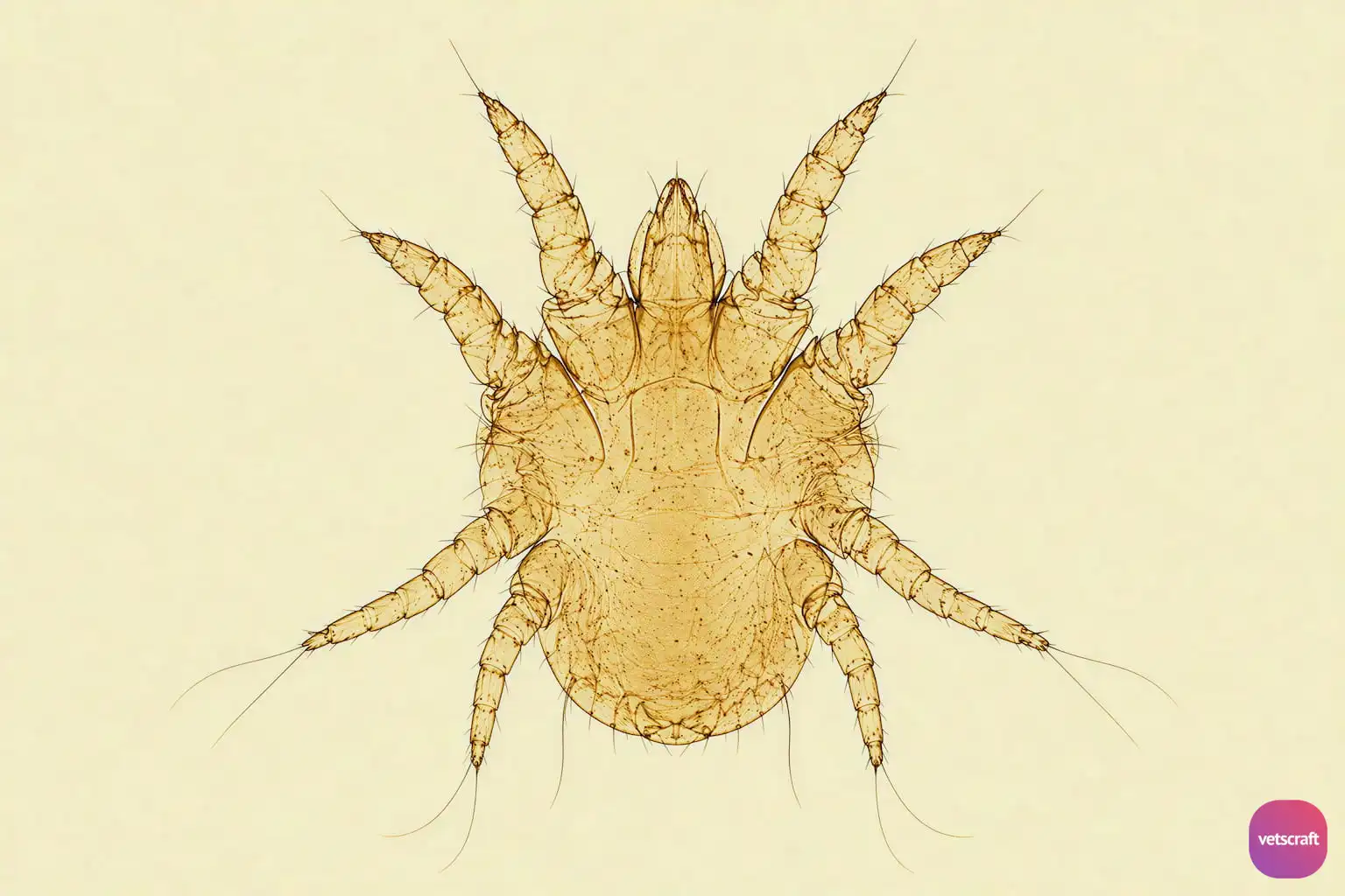
Morphology
- Non-burrowing mite, oval in shape.
- Has pointed mouthparts.
- All four legs are long and project well beyond the margin of the body.
- Tarsal suckers have three-jointed pedicels.
- Males have tarsal suckers on three-jointed pedicels on the first, second, and third pairs of legs.
- Females have suckers on three-jointed pedicels on the first, second, and fourth pairs of legs.
- The fourth pair of legs in males and the third pair of legs in females possess bristles.
- Males have a pair of copulatory suckers on the ventral aspect, as well as a pair of large terminal tubercles, each of which bears several hairs.
- The pubescent female is provided with a pair of posterior copulatory tubercles, which are absent in the ovigerous female, while the latter has a wide genital aperture on the anterior aspect of the ventral surface.
- The dorsal surface of the body is devoid of scales and spines, but the cuticle shows very fine striations.
- The posterior margin of the abdomen of the male is divided into two lobes.
Life Cycle
- The eggs are laid on the skin at the edges of the lesion and hatch in 1–3 days. If the eggs are separated from the skin, they take longer to hatch or may die.
- The larvae feed and molt to the nymphal stage in 2–3 days, spending 12 hours in a stage of lethargy.
- The nymphal stages last for 3–4 days, including a lethargic period of 36 hours before molting.
- Smaller nymphs usually become males.
- Pubescent females appear one day before males (as soon as 5 days after hatching in females and 6 days in males).
- Copulation begins soon after ecdysis and lasts one day. If there are more females than males, the period may be shorter. The male-to-female ratio is 1–2:4.
- The ovigerous female begins to lay eggs one day later, or nine days after hatching from eggs.
- The females live for 30–40 days and lay up to 90 or more eggs.
- Mites may survive for up to 2 weeks off the host.
- The life cycle period from egg to egg is about 12 days.
Pathogenesis
- The mites puncture the epidermis of the skin and suck lymph, stimulating local inflammatory swelling richly infiltrated with serum. The latter exudes onto the surface and coagulates, thus forming a crust and scab.
- The altered conditions cause the wool to become loose and fall out, or it is pulled out by the sheep. The sheep bite and scratch the lesions, which are markedly pruritic.
- The bare, crusty patches are unsuitable for the mites, which therefore migrate to the margins of the lesion and thus extend the process outward.
- A primary lesion can occur as early as two weeks after contact. This may be evident as a few strands of discolored wool, under which there is an area of moist dermatitis.
- In winter conditions, this can spread readily to large areas of the body. There is a type I hypersensitivity reaction by the host to antigenic material (feces) produced by the mites.
- Hypersensitivity causes inflammation, surface exudation, scales, and dry yellow crust formation, with excoriation due to self-trauma resulting in moist crusts.
Clinical Signs
- Scab lesions may occur on all parts of the body that are covered by wool or hair, but occur most frequently around the shoulder, along the sides of the body in wooled sheep, and along the back, sternum, and dorsal aspect of the tail in hairy sheep. It can affect all ages but is severe in young lambs.
- In early lesions, the wool is disturbed over the lesion by the biting and scratching of sheep and usually has a lighter color than the surrounding wool.
- A two- to four-day-old lesion appears as a small papule, about 5 mm in diameter, with a yellow and moist surface.
- From about the fifth day onward, the exudate begins to coagulate, forming pale yellow crusts, and the lesion extends outward as the number of parasites increases.
- Older lesions are easy to detect on account of the loss of wool and presence of scab, while mites are producing fresh foci in surrounding covered areas.
- If the wool is opened, it is matted together above the skin by scabs, underneath which numerous parasites are located. In latent cases, a small lesion may exist for months, seen as small, dry lesions on the scrotum, perineum, along the sheath, sternum, ear, infraorbital fossa, and at the base of the horns. Sheep scab is most active in autumn and winter, while latency tends to occur in summer. There is wool loss and weight loss in growing animals.
- When handled, infested sheep may demonstrate a “nibble reflex,” characterized by lip smacking and protrusion of the tongue; others may show fits lasting for 5–10 minutes. Secondary bacterial and fungal infections may also occur.
Diagnosis
It is simple to diagnose active disease in a scabby flock, but it is difficult to diagnose latent cases.
Diagnosis can be confirmed by microscopic examination of skin scrapings from suspected lesions.
Treatment
- Lime-sulfur dip.
- Deltamethrin (Butox) – 4 mL in one liter of water as a dip.
- Two injections of ivermectin at a dose rate of 200 µg/kg body weight by subcutaneous injection, 7 days apart.
- Moxidectin: 200 µg/kg body weight, subcutaneously; treatment regimens vary by formulation, with two injections commonly given 10 days apart.
- Doramectin: 300 µg/kg body weight, single dose subcutaneously, is effective.
- For ear canker/ear mange, topical application of acaricides with antibacterial compounds may be effective.
- Antihistamines, Vitamin A, D₃, E, and selenium supplementation may also be effective as supportive treatment. Concurrent treatment with antibiotics for secondary bacterial infection may also be useful.
Differences Between Sarcoptes and Psoroptes
| Sarcoptes sp. | Psoroptes sp. |
|---|---|
| Causes scabies in humans and may affect sheep, goats, cattle, pigs, equines, dogs, and rabbits; it is not host-specific. | Infests sheep, goats, cattle, dogs, and rabbits and is also host-specific. |
| Burrows into the skin and causes thickening of the skin. | Non-burrowing mite that causes scab formation. |
| Body is round. | Body is oval. |
| Legs are short. The anterior pairs may be visible outside the body, while the posterior two pairs are within the body. | Legs are long. All four pairs of legs project beyond the margin of the body. |
| Pedicel is unjointed and has bell-shaped suckers. | Pedicel is segmented and has funnel-shaped suckers. |
| The posterior part of the abdomen in males is not lobed. | The posterior part of the abdomen in males is bilobed. |
| In males and females, the tarsi of the 1st, 2nd, and 4th pairs and the 1st and 2nd pairs of legs, respectively, have suckers. | In males and females, the tarsi of the 1st, 2nd, and 3rd pairs and the 1st, 2nd, and 4th pairs of legs, respectively, have suckers. |

