TABLE OF CONTENTS
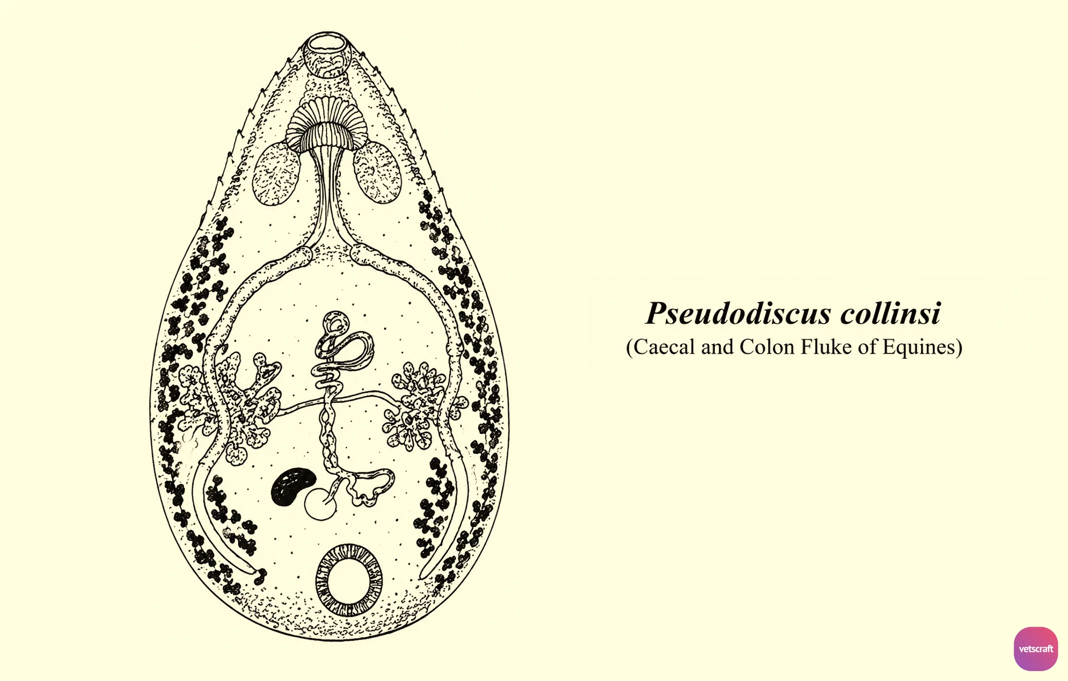
Pseudodiscus collinsi (Caecal and Colon Fluke of Equines)
Pseudodiscus collinsi is an amphistome trematode commonly found in the caecum and colon of equines. It is one of the important intestinal flukes reported in horses and can be identified by its characteristic body shape and reproductive organs. The parasite belongs to the family Gastrothylacidae and utilizes a freshwater snail as its intermediate host.
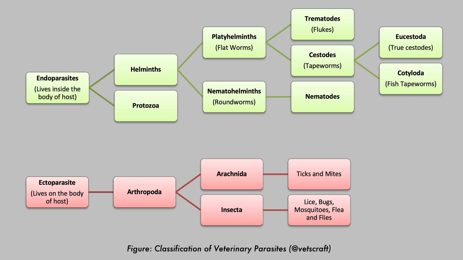
Taxonomical Classification
- Kingdom: Animalia
- Phylum: Platyhelminthes
- Class: Trematoda
- Subclass: Digenea
- Order: Echinostomida
- Suborder: Paramphistomata
- Superfamily: Paramphistomoidea
- Family: Gastrodiscidae
- Genus: Pseudodiscus
- Species: Pseudodiscus collinsi
Parasite Overview
- Common Name: Caecal/colon fluke of equines
- Host: Equines
- Prediction site / Location: Caecum and colon of equines
- Distribution: Commonly reported in horses in India
- Intermediate Host: Indoplanorbis exustus
Morphology
- Distinct anterior globoid and posterior discoid portions are absent.
- The body tapers anteriorly and is rounded posteriorly.
- An oral pouch is present.
- Intestinal caeca are wavy.
- Testes are deeply lobed and positioned side by side.
- Vitellaria are located mostly lateral to the intestinal caeca.
- The ovary is single, located posterior to the testes, and positioned laterally.
Life Cycle
Pseudodiscus collinsi has an indirect life cycle involving a freshwater snail as the intermediate host. Eggs passed in the feces of infected equines hatch in water and release miracidia, which penetrate suitable snail hosts. Within the snail, the parasite undergoes development through sporocyst, redia, and cercaria stages.
Cercariae leave the snail and encyst on aquatic vegetation as metacercariae. Equines become infected by ingesting vegetation contaminated with metacercariae during grazing. Following ingestion, immature flukes develop into adults and establish themselves in the cecum and colon, where they complete their life cycle.
For information on the general life cycle, epidemiology, pathogenesis, histopathological lesions, clinical signs, diagnosis, treatment, and control of amphistome infections, refer to our detailed article on Amphistomes. The principles discussed in that article are broadly applicable to infections caused by Pseudodiscus collinsi.