
Eimeria (Coccidia): Classification, Morphology, Oocyst Structure, and Life Cycle
Eimeria is a genus of obligate intracellular protozoan parasites belonging to the family Eimeriidae. These parasites are among the most important causes of coccidiosis in domestic and wild animals, particularly poultry, cattle, sheep, goats, and rabbits. Eimeria species are highly host-specific and primarily infect the intestinal epithelial cells, leading to enteritis, poor growth, reduced productivity, and, in severe cases, death.
Members of Eimeria have a direct (homoxenous) life cycle that includes asexual reproduction (schizogony or merogony), sexual reproduction (gametogony), and sporogony, which occurs outside the host. The infective stage is the sporulated oocyst, while the unsporulated oocyst is shed in the feces.
Taxonomical Classification of Eimeria
- Kingdom: Protista
- Phylum: Apicomplexa
- Class: Sporozoa
- Subclass: Coccidia
- Order: Eucoccidia
- Suborder: Eimeriina
- Family: Eimeriidae
- Genus: Eimeria
- Common Name: Coccidia
Members of the subclass Coccidia are typically intracellular parasites of vertebrates. They may be homoxenous or heteroxenous.
Members of the family Eimeriidae are homoxenous (single-host) intracellular parasites of intestinal epithelial cells. In their life cycle, both asexual and sexual reproduction occur in the same host.
Structure of Unsporulated Oocyst of Eimeria spp.
The fully matured oocyst breaks out of the host cell and is passed in the feces. The freshly passed oocyst is called an unsporulated oocyst, which contains an undifferentiated mass (single cell) called the sporont. The oocyst is the resistant stage of the life cycle.
Under favorable conditions, sporogony (often called sporulation) takes place in the environment. Following sporulation, the oocyst becomes infective to a susceptible host.
Structure of Sporulated Oocyst of Eimeria spp.
The oocysts are spherical, subspherical, ovoid, or ellipsoidal in shape. They vary in size from species to species. The oocyst wall is composed of two layers and is generally clear and transparent. However, it may be yellowish or even green in color in some species, while other species possess striations or punctuations in the wall. Several species of coccidia possess a micropyle at the pointed end, which may be covered by a dome-like structure called the micropyle cap.
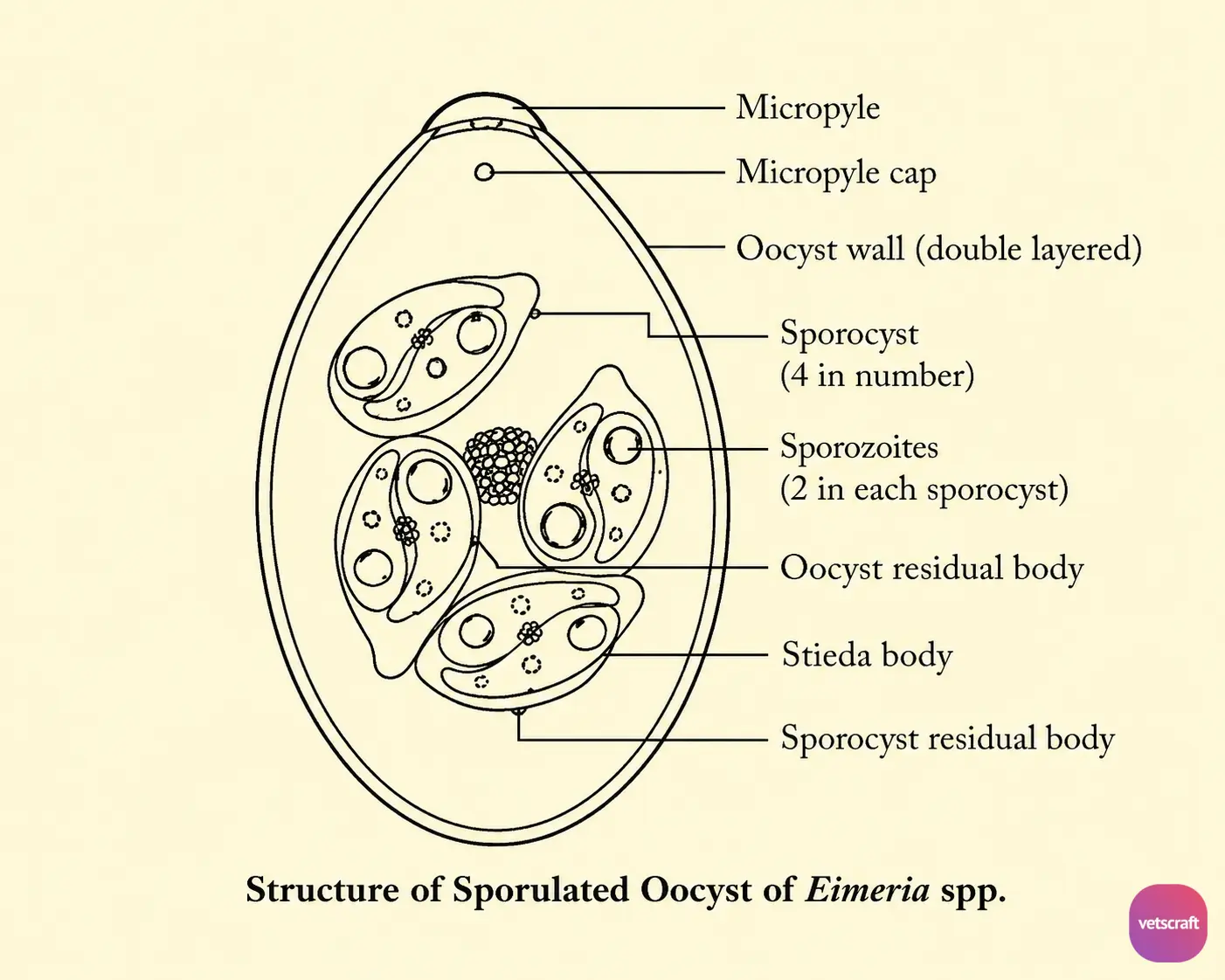
A sporulated oocyst of Eimeria spp. has four sporocysts, each containing two sporozoites. The sporocysts are more or less elongate or ovoid; one end is more pointed than the other, and a knob-like structure at the pointed end is called the Stieda body.
The sporozoites are usually elongate, with one end rounded and the other tapered (comma-shaped). They have a granular cytoplasm, a centrally placed nucleus, and homogeneous vacuoles at both ends called refractile bodies or globules.
An oocyst residual body and a sporocyst residual body may also be present in the oocyst. These are materials left over after the formation of the sporocyst and sporozoites, respectively.
General Life Cycle of Eimeria spp.
The definitive host (chickens, cattle, sheep, and goats) becomes infected by ingesting sporulated oocysts along with feed and water. Following ingestion, excystation occurs, and sporozoites are liberated from the oocyst in the intestine.
In large animals, excystation is facilitated by CO2, trypsin, and bile, whereas in chickens and other birds, the oocysts are mechanically ruptured in the gizzard.
The liberated sporozoites penetrate the intestinal wall at the tips of the villi, where they are engulfed by macrophages and carried to the glands of Lieberkühn. Soon after reaching the glands, the sporozoites leave the macrophages and enter the epithelial cells lining the glands to undergo further development.
Asexual Reproduction (Schizogony/Merogony)
The sporozoites that penetrate the epithelial cells round up and become trophozoites. The nucleus of each trophozoite divides by schizogony to form a schizont (meront) (first-generation schizont). This first-generation schizont produces about 900 merozoites (schizozoites) within 2–3 days of infection.
The mature schizonts rupture and release the first-generation merozoites into the lumen of the intestine. These first-generation merozoites then infect healthy epithelial cells and develop into second-generation schizonts (meronts). The second-generation schizont produces about 200–350 merozoites by the fifth day of infection.
Among the second-generation merozoites, the majority enter new cells and initiate the sexual phase of the life cycle. However, a small proportion of the merozoites continue asexual reproduction and produce third-generation schizonts.
Sexual Reproduction (Gametogony)
The majority of the second-generation merozoites (about 70%) destined for the sexual phase develop into macrogamonts within the epithelial cells, while a smaller number develop into microgamonts. Within each microgamont, a large number of tiny biflagellate microgametes are formed.
The microgametes break out of the host cell and enter cells containing macrogametes, where fertilization occurs, resulting in zygote formation. Thereafter, the macrogamete, containing plastin granules (wall-forming bodies), forms a thick wall around the zygote, called the oocyst wall.
The mature oocyst breaks out of the host cell and enters the lumen of the intestine, from where it is finally passed in the feces. The freshly passed oocyst contains an undifferentiated mass called the sporont.
Sporogony (Sporulation)
In Eimeria spp., sporogony takes place in the environment outside the host. During this process, the sporont divides into four sporoblasts, each of which forms a sporocyst containing two sporozoites. These sporulated oocysts are the infective stage for another susceptible host.

