
Chicken Coccidiosis: Causes, Eimeria Species, Life Cycle, Diagnosis, Treatment & Control
Chicken coccidiosis is one of the most economically important parasitic diseases of poultry worldwide. It is caused by protozoan parasites of the genus Eimeria, which invade the intestinal tract and cause enteritis, poor growth, reduced feed efficiency, decreased egg production, and mortality in severe cases. The disease is primarily transmitted through ingestion of sporulated oocysts contaminating feed, water, or litter.
Among the seven Eimeria species affecting chickens, E. tenella, E. necatrix, E. maxima, E. acervulina, and E. brunetti are the most pathogenic. This article provides a comprehensive overview of chicken coccidiosis, including its causes, classification, Eimeria species, life cycle, pathogenesis, clinical signs, diagnosis, treatment, anticoccidial drugs, vaccines, immunity, and control measures.
Chicken coccidiosis is one of the most important diseases of chickens, causing significant economic losses due to mortality, reduced weight gain, and decreased egg production. The disease is transmitted through feed and water contaminated with sporulated oocysts. Because of its ubiquitous nature, even birds maintained in cage systems may become infected.
In caged housing systems, houseflies and rodents are suspected to play a role in contaminating feed and water with coccidian oocysts. This may be one of the main reasons for the occurrence of coccidiosis in caged layers and broilers. Coccidiosis is generally a self-limiting disease.
Seven species of Eimeria are known to infect chickens, of which E. tenella, E. necatrix, E. maxima, E. brunetti, and E. acervulina are the most important and pathogenic species in domestic chickens. Mixed infections are common under natural conditions.
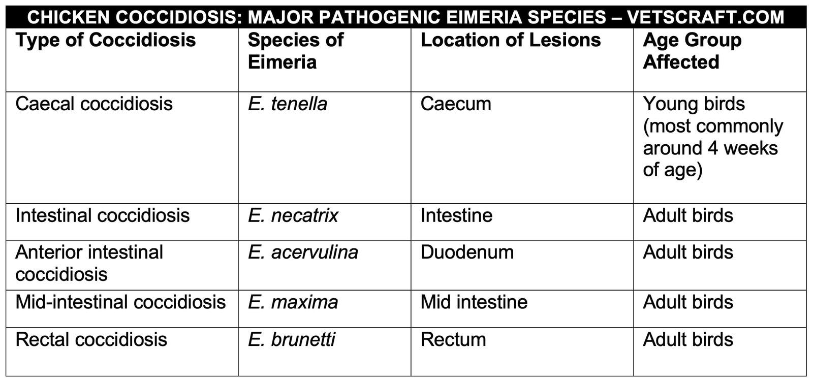
Eimeria species affecting chickens are highly host- and site-specific. Therefore, based on the type of lesions they produce, chicken coccidiosis is classified as follows:
Caecal Coccidiosis (Eimeria tenella)
Caecal coccidiosis is caused by Eimeria tenella, the most common and pathogenic coccidian species affecting domestic chickens.
- Location: All developmental stages occur in the caecum.
- Age: Caecal coccidiosis is most frequently seen in young birds.
Oocyst Morphology
- Ovoid, 14–31 × 9–25 µm
- Oocyst wall smooth
- No micropyle
- No residuum
- Polar granule present
Sporulation Time
18 hours at 29°C and 24–48 hours at room temperature. However, sporulation does not occur below 8°C.
Epidemiology
Caecal coccidiosis primarily affects young birds. Chicks around 4 weeks of age are the most susceptible, whereas chicks 1–2 weeks old are relatively resistant to infection. Similarly, older birds are generally immune as a result of previous exposure.
Caecal coccidiosis is more common in broilers. Under field conditions, all birds are exposed to infection, but clinical coccidiosis occurs only when large numbers of oocysts are ingested within 72 hours. In contrast, if birds ingest small numbers of oocysts over a prolonged period, immunity develops rather than clinical coccidiosis.
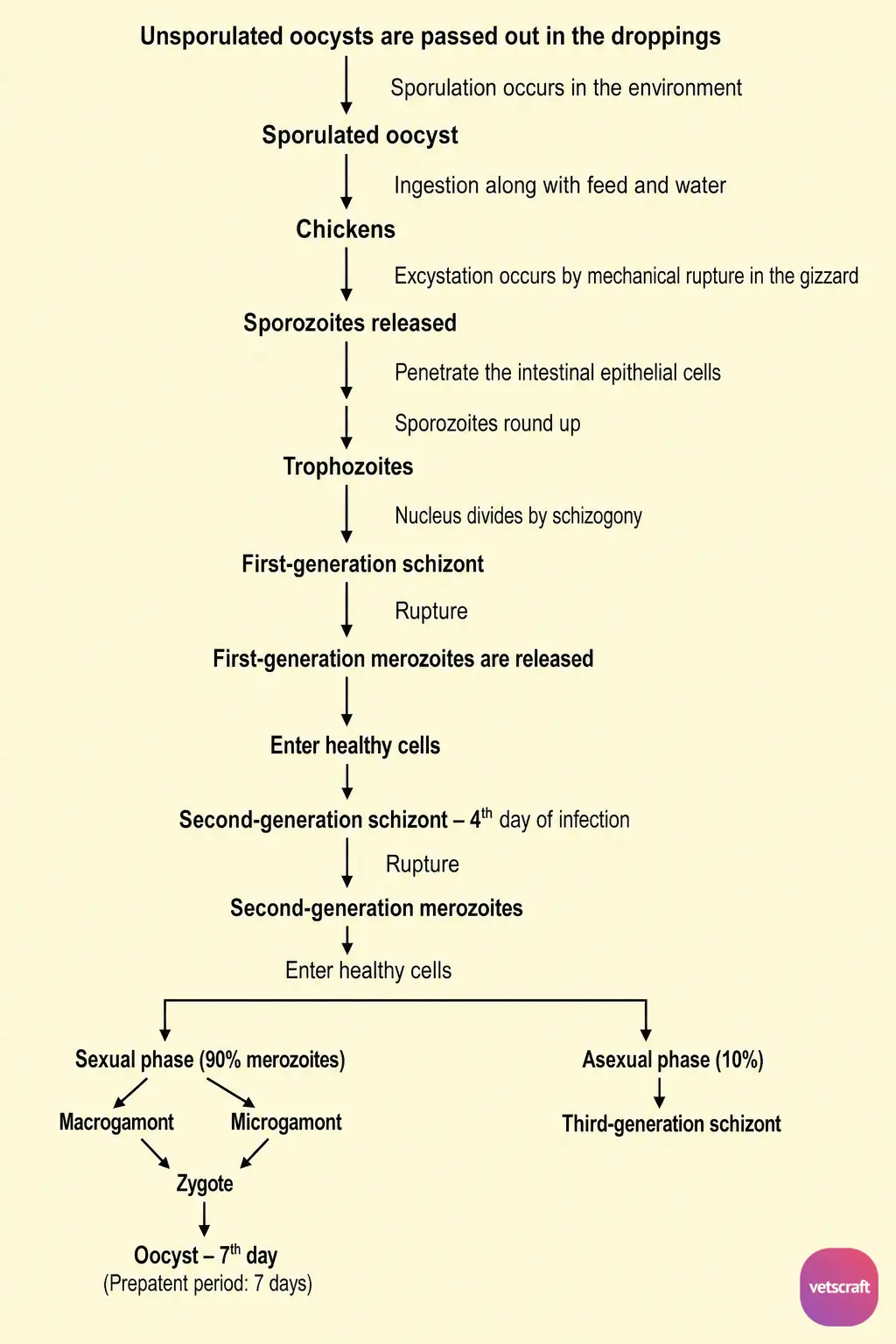
Life Cycle of E. tenella
Pathogenesis and Clinical Signs
The clinical manifestations of caecal coccidiosis may vary from an inapparent infection to an acute, highly fatal disease. The severity of the disease depends on the dose of oocysts, the strain of Eimeria spp. involved, breed, age, nutritional status, concurrent infections, and stress.
The first signs of the disease may appear within 72 hours of infection, usually coinciding with the development of second-generation schizonts, which are primarily responsible for the pathogenesis.
The large second-generation schizonts may cause rupture of delicate capillaries, resulting in blood in the droppings by the fourth day of infection. Severe hemorrhage usually occurs on the fifth or sixth day of infection; therefore, mortality is generally highest between the fifth and sixth days. By the eighth or ninth day, affected birds either die or recover.
Birds that recover from the acute disease may continue to suffer from chronic illness due to a persistent caecal core. Under the normal course of the disease, the caecal cores are expelled approximately 14 days after infection.
Clinical signs of caecal coccidiosis include drooping of the wings and head, listlessness, bloody diarrhea, huddling together to keep warm, and cessation of feed intake, while water consumption usually remains normal.
Postmortem Lesions
The lesions of caecal coccidiosis observed on different days after infection are as follows:
- Third Day: Petechial hemorrhages
- Fourth Day: Numerous hemorrhagic spots appear
- Fifth Day: Dilation of the caeca
- Sixth Day: Intestinal contents contain unclotted or partially clotted blood, schizonts, and merozoites.
- Seventh Day: Gamonts can be seen in mucosal scrapings. The caecal contents become consolidated, caseous, and adherent to the mucous membrane.
- Eighth Day: The lumen of the caecum is filled with a caseous plug.
- Ninth Day: The caecal core detaches from the mucous membrane.
- Tenth–Fourteenth Day: The caecal core is passed in the droppings.
Immunity
Coccidiosis is a self-limiting disease in the absence of reinfection. Recovered birds develop immunity to reinfection with E. tenella but remain susceptible to other Eimeria species. Second-generation schizonts are primarily responsible for inducing immunity. Concurrent immunosuppressive diseases, such as infectious bursal disease (IBD) and Newcastle disease (ND), increase the severity and mortality associated with coccidiosis in chickens.
Intestinal Coccidiosis (Eimeria necatrix)
Intestinal coccidiosis is caused by Eimeria necatrix, the second most pathogenic species of Eimeria. It is an important cause of chronic coccidiosis in layer chickens and produces permanent scarring of the intestine.
The biotic potential of this species is low, meaning it produces relatively few oocysts. Therefore, a longer period is required to contaminate the environment with large numbers of oocysts.
- Location: First- and second-generation schizogony (asexual reproduction) occur in the small intestine, while gametogony (sexual reproduction) occurs in the caecum.
- Age: Older birds, generally 9–14 weeks of age, are affected.
Oocyst Morphology
- Ovoid, 12–29 × 11–24 µm
- Oocyst wall smooth and colorless
- No micropyle
- No residuum
- Polar granule present
Sporulation Time: 18 hours to 2 days.
Life Cycle
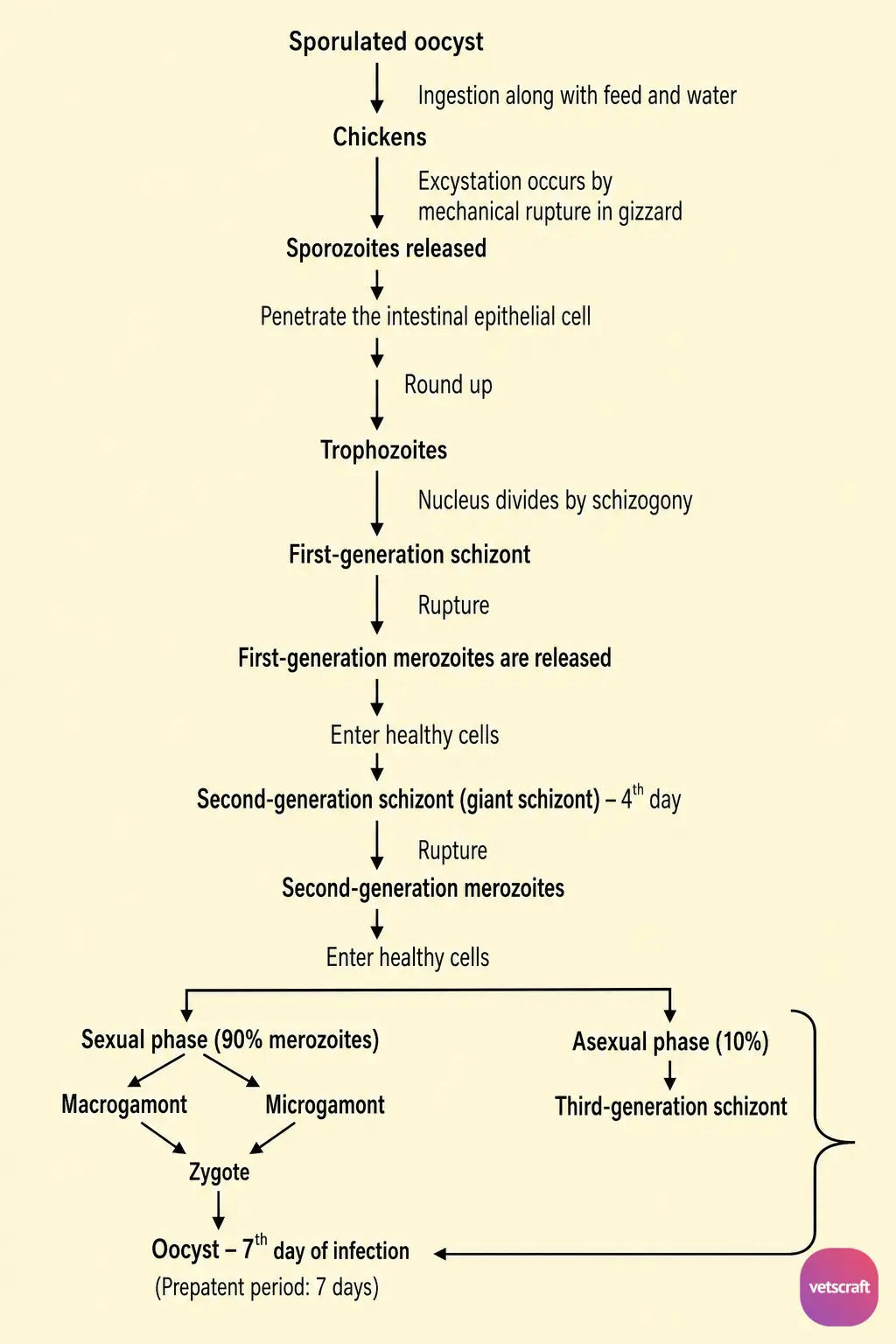
The life cycle is similar to that of E. tenella. However, first- and second-generation schizogony (asexual reproduction) occur in the small intestine, whereas third-generation schizogony and gametogony (sexual reproduction) occur in the caecum.
Pathogenesis
Intestinal coccidiosis is usually manifested as a chronic disease in older birds, especially layer pullets 9–14 weeks of age. The severity of the disease depends on the number of oocysts ingested and the level of immunity resulting from previous light infections.
Postmortem Lesions
The lesions are mainly observed in the middle third of the small intestine on different days after infection:
- Fourth Day: Developing second-generation schizonts in the deeper layers of the intestine appear as small, white, opaque foci in the submucosa. These are visible from the serosal surface but not from the mucosal surface.
- Fifth–Sixth Day: Severe hemorrhage occurs due to the large, deeply located second-generation giant schizonts. The small intestine becomes markedly swollen (ballooned) and filled with unclotted blood. The intestinal wall is thickened, dull red, and gangrenous, and the intestine loses its contractile ability.
- The deeply located white, opaque schizont foci, together with the surrounding hemorrhagic areas, produce the characteristic “salt and pepper” appearance.
- End of the Sixth Day: Sloughing of epithelial cells occurs. The affected areas become infiltrated with fibrin and mononuclear cells, which are subsequently replaced by connective tissue. This leads to the formation of permanent scars that persist for a long time.
Death usually occurs 5–7 days after infection. The caeca are not severely affected, although they may become contracted and their contents dehydrated. Occasionally, blood may be present in the caeca due to excessive hemorrhage, which can lead to confusion with caecal coccidiosis.
Even after recovery, affected birds remain emaciated for several weeks. Therefore, retaining birds affected with intestinal coccidiosis is generally not considered economically worthwhile.
Mid-Intestinal Coccidiosis (Eimeria maxima)
Eimeria maxima causes mid-intestinal coccidiosis.
Oocyst Morphology
- Large, ovoid, approximately 29 × 23 µm.
- Oocyst wall is slightly yellow; some oocysts may have a roughened surface.
- No micropyle.
- No residuum.
- Polar granule present.
- Sporulation Time: 30 hours to 2 days.
Life Cycle
All developmental stages occur in the small intestine. The second- and third-generation schizonts are relatively small, whereas the gamonts are larger and are primarily responsible for the pathogenesis.
Pathogenesis
This species is moderately pathogenic. However, in severe infections, numerous petechial hemorrhages and marked mucus production occur in the intestine. The intestinal mucosa becomes inflamed and thickened, and as it loses its tone, it becomes flaccid and dilated. The intestinal contents are typically viscid, pinkish, mucoid, and yellow-orange in color.
Immunity
Infections with this species induce immunity rapidly, leading to early termination of the infection. Because E. maxima is highly immunogenic, its developmental stages are used in the production of coccidial vaccines.
Anterior Intestinal Coccidiosis (Eimeria acervulina)
Eimeria acervulina causes anterior intestinal coccidiosis. This species is less pathogenic but has a very high biotic potential; therefore, it produces large numbers of oocysts. It produces characteristic ladder-like lesions in the anterior third of the intestine.
Rectal Coccidiosis (Eimeria brunetti)
Eimeria brunetti causes rectal coccidiosis. The lesions produced by this species appear as white, bran-like deposits in the rectal region. Its oocysts do not sporulate under laboratory (in vitro) conditions.
Diagnosis
- The best method for diagnosing chicken coccidiosis is postmortem examination of representative birds from a suspected flock to identify the characteristic lesions.
- Examination of Intestinal Scrapings: Examination of intestinal scrapings helps diagnose chicken coccidiosis if birds die during the incubation period. Various developmental stages, such as schizonts and gamonts, can be demonstrated.
- Direct Microscopic Examination of Scrapings: Place a small amount of scraped material on a glass slide, add a few drops of tap water, cover with a coverslip, and examine under a microscope at 10× and 45× magnification.
- Examination of Intestinal Smear: Place the scraped material on a glass slide, prepare a smear, and allow it to air-dry. Stain the smear with Giemsa or Leishman stain using the same procedure as for a blood smear. After staining, examine under a microscope at 100× magnification, where schizonts and gamonts can be observed.
- Clinical Signs: Hemorrhagic diarrhea with bloody mucus, droopiness, and huddling together.
- Fecal Examination: Fecal examination is not reliable because the presence or absence of oocysts does not necessarily indicate clinical coccidiosis. For example, in E. tenella infection, the major pathogenic changes occur before oocysts are shed in the feces. Similarly, during the early stage of infection, oocysts may not be detected in the feces.
- Likewise, the number of oocysts per gram of feces does not accurately reflect the severity of the disease because it depends on the biotic potential of the species. For example, E. necatrix produces relatively few oocysts due to its low biotic potential but causes severe disease, whereas E. acervulina produces large numbers of oocysts because of its high biotic potential but is comparatively less pathogenic.
- Feces can be examined by direct microscopic examination, concentration sedimentation, or flotation techniques. Saturated salt solution, sugar solution, and Sheather’s solution are commonly used for flotation of oocysts.
- Histopathological Examination of the Intestine/Caecum: Performed to demonstrate the developmental stages of the parasite.
- Immunodiagnosis: IFA, IHA, ELISA, and DOT-EIA.
In Vitro Sporulation
Coccidiosis-positive fecal samples should be subjected to in vitro sporulation in the laboratory to identify Eimeria species based on the time required for sporulation.
Procedure
- Emulsify the coccidiosis-positive fecal sample by adding a sufficient quantity of 2.5% potassium dichromate solution.
- Pour the emulsified suspension into a Petri dish and cover it with a lid, leaving a small gap for aeration.
- Leave it at room temperature for 2 days. During this period, the dish may be gently rotated occasionally to facilitate aeration and ensure uniform sporulation.
Treatment
Coccidiosis can be controlled by treating affected birds with therapeutic drugs and prevented by the use of prophylactic drugs and vaccines. The drugs used for treatment are broadly classified as coccidiostatic and coccidiocidal.
The following drugs are commonly used:
| S. No. | Class and Name of the Drug | Mode of Action | Dose and Mode of Administration |
|---|---|---|---|
| I. | Sulphonamide Group 1. Sulphadimidine | Structural antagonist of para-aminobenzoic acid (PABA). It inhibits the conversion of dihydrofolic acid to tetrahydrofolic acid, which is required for DNA synthesis. It acts on first- and second-generation schizonts. At low doses, it acts as a coccidiostat, whereas at high doses it acts as a coccidiocide. |
0.4% in feed and 0.2% in water. Treatment Course 3 days of treatment → 2-day interval → 3 days of treatment. |
| 2. Sulphaquinoxaline |
0.043% in water. Treatment Course 2 days of treatment → 3–5 day interval → 2 days of treatment. | ||
| II. | Thiamine Analogues 1. Amprolium | 0.0125% | |
|
2. Sulphaquinoxaline + Amprolium This combination is more effective. | 0.006% in feed | ||
| 3. Amprolium + Ethopabate | 250–500 g/tonne of feed | ||
| III. | Nitrobenzamides 1. Zoalene | Prophylactic | 0.01–0.015% in feed |
| 2. Nitromide | 0.1% in feed | ||
| 3. DOT (Dinitro-O-Tolumide) | 0.5 kg/tonne of feed. It is commonly used as an anticoccidial in broiler starter feed. | ||
| IV. | Nitrofurans 1. Nitrofurazone | Acts on second-generation schizonts of E. tenella and E. necatrix. | 0.022% |
| 2. Furazolidone | Broad-spectrum antibacterial and coccidiostatic activity. | 0.0055% in feed | |
| 3. Nitrofurazone + Furazolidone Combination | Produces a marked coccidiostatic effect. | 0.0053% + 0.0008% | |
| V. | Substituted Carbanilides 1. Nicarbazin | 0.0125% in feed | |
| VI. | Ionophore Antibiotics (Fermentation products of Streptomyces albus) 1. Monensin |
Ionophores act on the intestinal free stages of coccidia (sporozoites, merozoites, and gametocytes). Monovalent ionophore: Binds monovalent ions (Na+ and K+). | 0.01% |
| 2. Lasalocid | Divalent ionophore: Binds divalent ions (Ca2+ and Mg2+). | 0.005–0.0075% | |
| 3. Salinomycin | Monovalent ionophore. | 0.01% | |
|
4. Maduramicin (Fermentation product of Actinomadura yumaensis) | Monovalent ionophore. | 5–6 ppm | |
| VII. | Recent Drugs 1. Sulphachloropyrazine sodium (ESB3) | 1–5 g/L drinking water | |
| 2. Toltrazuril | 75 ppm in feed | ||
| 3. Diclazuril | 75 ppm in feed | ||
| 4. Clopidol | 0.5 kg/tonne of feed |
Anticoccidial Resistance
Continuous and indiscriminate use of anticoccidial drugs may result in the development of drug-resistant strains. Once resistance develops, the recommended dose that was previously effective is no longer sufficient, leading to the use of higher drug doses to control coccidiosis. This not only increases production costs but also creates additional problems such as drug residues in meat and eggs.
To minimize the development of anticoccidial resistance under field conditions, different classes of anticoccidial drugs should be used in rotation rather than relying on a single drug for prolonged periods. The following programs are commonly practiced under field conditions to reduce resistance.
Straight Program
This method is adopted in broiler production, in which the same ionophore anticoccidial is added to both starter and grower feeds. The concentration of the drug may be increased in the grower feed to provide maximum protection during 3–4 weeks of age, the period of greatest susceptibility to caecal coccidiosis. This is known as the step-up program. Alternatively, the concentration of the drug may be reduced in the grower or finisher feed, which is known as the step-down program.
Shuttle Program
This method is practiced in layer production, in which a chemical anticoccidial is added to the starter feed and an ionophore anticoccidial is added to the grower feed. This strategy helps minimize the development of resistance because the duration of exposure to the same drug is limited.
Control of Coccidiosis in Chickens
Control of coccidiosis in chickens includes the following:
- Immunoprophylaxis
- Precocious Line
- Control of Coccidiosis by Management
1. Immunoprophylaxis
| Type of Vaccine | Name of the Vaccine / Contents | Mode and Age of Vaccination |
|---|---|---|
| I. Non-attenuated Vaccine (Live) |
1. Coccivac-D Live virulent oocysts of E. acervulina, E. brunetti, E. maxima, E. mitis, E. necatrix, E. praecox, and E. tenella. | Used in layers and breeders. Birds are vaccinated at 3 days of age, followed by low-level anticoccidial medication starting at 13 days of age and continuing until 6 weeks of age. |
|
2. Coccivac-B Live virulent oocysts of E. acervulina, E. maxima, E. mitis, and E. tenella. | Used in broilers. | |
|
3. Immucox Chickens-I Virulent oocysts of all species except E. brunetti. | Used in broilers. | |
| 4. Immucox Chickens-II | Used in layers and breeders. | |
| II. Attenuated Vaccine |
1. Paracox Attenuated precocious-line strains of all Eimeria species. | |
| 2. Livacox-T | ||
| 3. Livacox-D | ||
| III. Subunit Vaccine |
CoxAbic The first subunit vaccine developed against coccidiosis. It contains purified gametocyte proteins of E. maxima. |
Used in broilers. Breeder birds are vaccinated twice before the onset of laying. First dose: 12–16 weeks of age. Second dose: 18–20 weeks of age (0.5 mL per dose). The antibodies produced are transferred to chicks through the yolk, providing protection for up to 6 weeks. |
2. Precocious Line
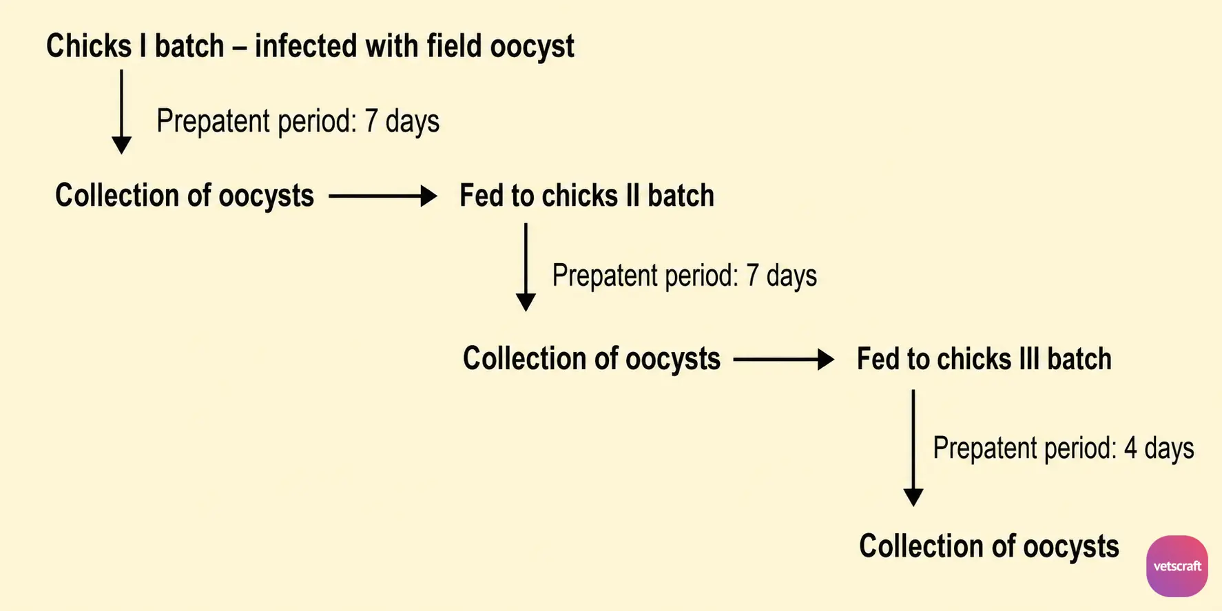
Oocysts obtained during a shortened prepatent period by bypassing second-generation schizogony are referred to as a precocious line. In these parasites, the first-generation merozoites differentiate directly into gametes, which subsequently fuse to form zygotes, resulting in oocyst production within approximately 4 days.
Development of Precocious Line
Chicks are repeatedly infected with a virulent strain, and the first oocysts produced are collected. After repeated passages, the oocysts gradually lose their virulence while retaining their immunogenicity. These are known as precocious oocysts and can be used for vaccine production.
3. Control of Coccidiosis by Management
- Destroy oocysts by fumigation with ammonia or methyl bromide.
- Heap litter to generate heat, which helps destroy oocysts.
- Raise young birds separately.
- Keep feeders and waterers clean.
- Raise chicks in cage systems.
- Maintain proper sanitation and good management practices.
- Control flies, rats, and mice.
- Avoid dampness and water stagnation in and around the farm.
- Separate affected birds from healthy birds and treat them promptly.
- Administer a coccidiostat to healthy birds as a preventive measure.
- Avoid water spillage.
- Avoid feed ingredients that promote wet litter conditions.
- Burn dead birds for proper disposal.
- Keep litter dry and stir it frequently.
- Maintain proper lighting and avoid overcrowding.
- Anticoccidial drugs may be administered before or after debeaking, deworming, and vaccination.

