TABLE OF CONTENTS
Blood Flukes (Schistosomatidae): Morphology, Life Cycle, Pathogenesis, Treatment and Control
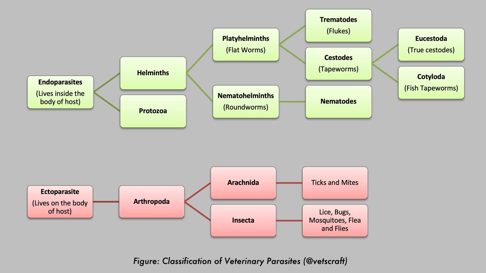
Blood flukes belonging to the family Schistosomatidae are important trematode parasites that inhabit the blood vessels of domestic and wild animals. Unlike most flukes, schistosomes are dioecious (unisexual) and exhibit marked sexual dimorphism. Species such as Schistosoma nasale, Schistosoma spindale, Schistosoma indicum, and Schistosoma incognitum are of major veterinary importance because they cause schistosomiasis, nasal granuloma, and associated vascular lesions in livestock.
Blood flukes are found in blood vessels; hence, they are called “blood flukes”. They are elongate, cylindrical, and resemble roundworms. They are unisexual and exhibit marked sexual dimorphism.
The pharynx is absent. The digestive system begins with an oral opening leading to the esophagus, and the intestine divides into two branches at the level of the ventral sucker.
The branched caeca unite to form a common caecum. The oral sucker and ventral sucker are very close to each other. The suckers are not very strong.
Taxonomical Classification
- Kingdom: Animalia
- Phylum: Platyhelminthes
- Class: Trematoda
- Subclass: Digenea
- Order: Strigeidida
- Family: Schistosomatidae
- Genus: Schistosoma
Parasite Overview
- Common Name: Blood Flukes
- Family: Schistosomatidae
- Major Diseases: Schistosomiasis, Nasal Schistosomiasis (Nasal Granuloma or Snoring Disease), and Cercarial Dermatitis (Swimmer’s Itch)
- Definitive Hosts: Cattle, buffaloes, sheep, goats, pigs, dogs, horses, and other mammals (depending on the species)
- Intermediate Hosts: Freshwater snails such as Indoplanorbis exustus and Lymnaea luteola
- Site of Predilection: Blood vessels, particularly the nasal, mesenteric, and portal veins
- Infective Stage: Cercaria
- Diagnostic Stage: Eggs in nasal discharge or feces
- Mode of Transmission: Penetration of cercariae through the skin or mucous membranes during contact with contaminated water
- Metacercarial Stage: Absent
- Unique Feature: Schistosomes are dioecious (unisexual) trematodes with marked sexual dimorphism
Morphology
Morphology of Male
- Males are short and stumpy. The cuticle has tuberculations in S. nasale, whereas in S. spindale it is smooth.
- The lateral edges of the body on the ventral aspect of the male curve inward and form a gutter-like groove known as the gynaecophoric canal. During copulation, the female fluke is lodged within this groove.
- Testes number 4 to 6, although they may exceed 60 in some species.
- The genital pore opens posterior to the ventral sucker.
Morphology of Female
- Females are slender and longer than males. The ovary is single and compact, situated anterior to the common caecum.
- The vitelline gland occupies the region posterior to the ovary.
- After copulation, females leave the male and migrate to their respective sites to lay eggs.
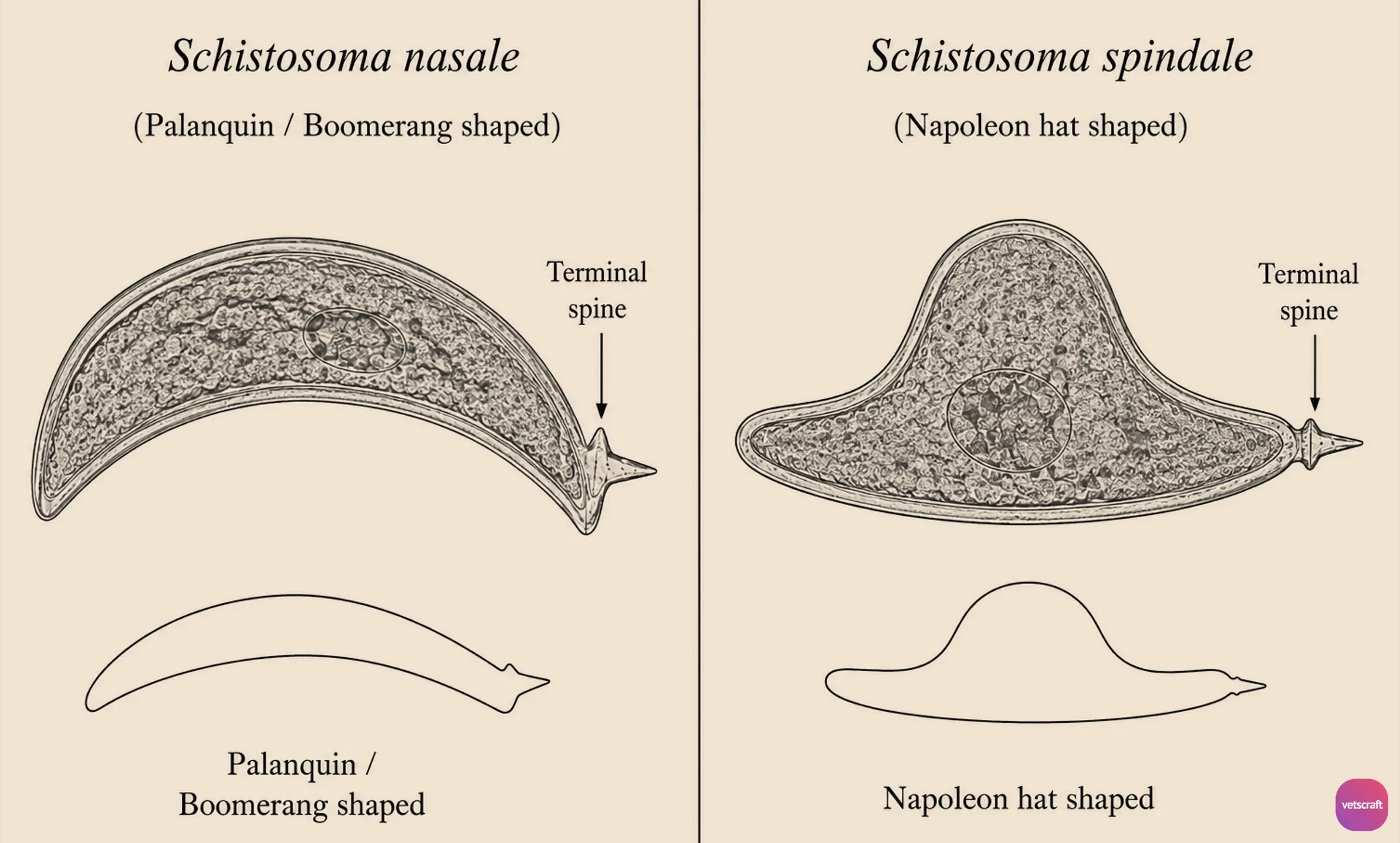
- The eggs are thin-shelled and non-operculated; in some species, they possess a lateral or terminal spine.
Comparative Morphology of Schistosomes
| Character | S. nasale | S. spindale | S. indicum | S. incognitum |
|---|---|---|---|---|
| Definitive Hosts | Cattle and buffaloes usually remain symptomless (more common in bullocks); rarely found in sheep and goats | Ruminants and dogs | Discovered by British scientist R. E. Montgomery in 1906 from a horse. Occurs in all domestic animals except pigs. | Pigs and dogs |
| Intermediate Hosts | Indoplanorbis exustus | Indoplanorbis exustus | Indoplanorbis exustus | Lymnaea luteola |
| Cuticle | Tuberculated | Smooth | Finely tuberculated | Moderately tuberculated |
| Eggs | Palanquin/Boomerang-shaped | Napoleon hat-shaped | Oval-shaped | Oval-shaped |
| Testes | 2–4 | 3–7 | 5–12 | 2–7 |
| Site of Predilection | Nasal veins | Mesenteric and portal veins | Mesenteric and portal veins | Portal vein |
Note: Schistosoma bovis occurs in ruminants, horses, and humans and is found in the mesenteric and portal veins.
Life Cycle
The eggs are palanquin/boomerang-shaped and contain a fully developed miracidium with a terminal spine. Females lay eggs in the nasal veins. With the help of the terminal spine, the eggs penetrate the vein wall and reach the nasal passages, from where they are expelled in the nasal discharge.
The eggs hatch upon contact with water and release miracidia. The miracidium has a blunt anterior end, a pair of eyespots, and penetration glands. It also possesses numerous germ cells.
The miracidium enters the aquatic snail (Indoplanorbis exustus) by penetration and transforms into a first-generation sporocyst, which in turn produces daughter sporocysts.
The redial stage is absent. Numerous cercariae develop within the daughter sporocysts.
The cercariae actively emerge from the snail and swim in water for 24 to 48 hours, after which they die.
Emergence of cercariae from the snail is periodic and commonly occurs between 10:00 and 14:00 hours during the daytime; however, it depends on the environmental temperature.
Description of Cercaria
- Furcocercous cercaria, apharyngeate, and brevifurcate (short).
- The cercaria has 3 pairs of flame cells and 3 to 5 pairs of penetration glands.
- Cystogenous glands are absent; therefore, they do not encyst.
- Infection of the definitive host occurs by penetration of cercariae through the legs, oral mucosa, or nasal mucosa when animals come into contact with or drink contaminated water.
- The metacercarial stage is absent.
- Following penetration, the cercariae lose their tails and transform into schistosomula (young stage).
- The schistosomula are transported through various vital organs via the circulation and finally enter the systemic circulation, reaching their respective predilection sites within 4 to 7 days after infection.
- The life cycle of S. bovis, S. spindale, S. indicum, and S. incognitum (or S. suis) is similar to that of S. nasale, except that the eggs are voided in the feces and schistosomula are found in the portal vein from the 8th day after infection.
- Copulation occurs in the portal vein, and the flukes subsequently migrate to the mesenteric veins to mature and lay eggs.
Pathogenesis
Pathogenesis mainly occurs due to the following four stages:
- Due to invading cercariae
- Due to schistosomula
- Due to adults
- Due to eggs
1. Due to Invading Cercariae
Cercariae cause “cercarial dermatitis”, “swimmer’s itch”, “clam-digger’s itch”, “rice paddy itch”, or “lake-side disease” when they accidentally penetrate the skin of an unnatural host (humans).
2. Due to Schistosomula
Schistosomula are transported through various organs before reaching their predilection sites.
While passing through the lungs, they cause pneumonia. In other organs such as the pancreas and brain, they cause inflammation, hemorrhage, and congestion. In heavy infections, schistosomula may obstruct capillaries.
3. Due to Adults
In S. nasale, the presence of tuberculations causes constant irritation of the nasal veins, resulting in phlebitis.
It also causes cellular infiltration, thrombus formation, fibrous tissue proliferation, and thickening of the vessels, leading to stagnation of blood flow and rupture of capillaries.
Hemorrhage occurs due to the blood-feeding activity of the parasites, resulting in dilation of vessels and thrombus formation within veins.
4. Due to Eggs
The pathogenic effects caused by eggs are of major importance in schistosome infections. Eggs with terminal spines pierce capillaries and pass into the surrounding tissues, where they release toxins that induce chronic inflammation, leading to “Actinobody” or “Pseudotubercle” formation (Granuloma/Hoeppli’s reaction). This process occurs in three stages:
Stage of Cellular Infiltration
During this stage, phagocytic cells such as macrophages and neutrophils, followed by mononuclear cells, are attracted toward the eggs and accumulate around them.
Stage of “Actinobody” Formation
During this stage, the live miracidium releases toxins or lytic substances through pores in the eggshell. These lytic substances dissolve the invading cells in a radial pattern.
Stage of Abscess Formation
The stage of abscess formation occurs due to the destruction of invading cells by lytic substances. The abscess may rupture, resulting in ulcer formation in the intestine or nasal mucosa. The ulcer is subsequently covered by a granulomatous reaction. Papillomatous or cauliflower-like growths may also develop, giving the surface a rough appearance.
In chronic infections, fibrous tissue proliferation and thickening of the nasal or intestinal mucosa occur, depending on the species of schistosome involved. Occasionally, eggs are carried through the circulation and reach vital organs such as the liver, spleen, kidneys, and brain, where they cause fibrous encapsulation and calcification. Calcification and fibrous encapsulation in the brain may result in neurological signs.
Symptoms
Clinical Symptoms in S. nasale
The disease is common in working cattle. Clinical signs include rhinitis, mucopurulent nasal discharge containing blood, sneezing, coryza, dyspnea, respiratory distress, snoring, and nasal mucosa studded with tiny nodules. A whistling sound may be heard due to narrowing of the nasal passages. A cauliflower-like sessile growth develops in the nasal cavity and is known as a “nasal granuloma”. Feed intake and working capacity of the animal are reduced, and affected animals become easily fatigued.
As buffaloes are natural hosts, the pathogenesis is generally less severe. Only pinhead-sized nodules and congestion of the mucous membranes are usually observed.
Clinical Symptoms in S. spindale
Acute heavy infection is manifested by profuse diarrhea or dysentery, along with abscesses and ulcers in the intestinal mucosa. Anorexia and dehydration may also be present.
In chronic cases, anemia, emaciation, eosinophilia, hypoalbuminemia, hypergammaglobulinemia, and sometimes edema may occur. Neurological symptoms may also be observed.
Treatment
- Sodium antimony tartrate: 1.5 mg/kg body weight (IV), twice daily for 2 days.
- Potassium antimony tartrate (Tartar emetic): 2 mg/kg body weight (IV).
While administering the above two drugs, care should be taken because extravasation may cause cellulitis (phlegmon). Therefore, the drugs should be administered diluted in normal saline or dextrose saline.
- Lithium antimony thiomalate (Anthiomaline): Drug of choice. It should be administered in three divided doses of 20, 15, and 15 mL at 2-day intervals by deep IM injection.
Precautions during Anthiomaline drug administration: Injections should be given on alternate sides and the injection site should be massaged thoroughly. Avoid deposition of the solution beneath the skin, and do not administer Anthiomaline intravenously.
- Antimoson: 5 mL IM. The dose may be repeated after 4 days.
- Praziquantel: 60 mg/kg body weight (oral), effective in sheep and goats. It should be administered as three doses of 20 mg/kg at 4-hour intervals.
- Niridazole: 25 mg/kg body weight (oral), reported to be highly effective against S. suis.
- Triclabendazole: 20 mg/kg body weight.
Control
- Restrict animal access to lakes, ponds, and other stagnant water bodies.
- Provide a safe piped or well-water supply.
- Animals should not be allowed to drink from contaminated water sources.
- Snail populations may be reduced by increasing water flow in irrigation channels, which can wash snails downstream.
- Store water for at least 48 hours before offering it to animals, as cercariae will die during this period.
- Identify and treat infected animals promptly.