TABLE OF CONTENTS
Intestinal or Visceral Schistosomosis in Animals: Symptoms, Diagnosis, Treatment & Control
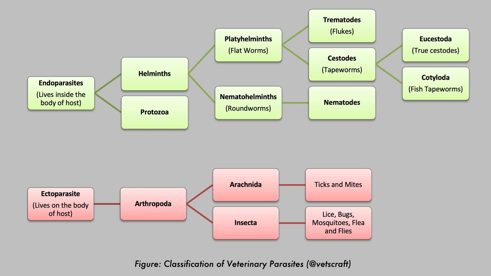
Intestinal or visceral schistosomosis is a parasitic disease of domestic animals caused primarily by blood flukes such as Schistosoma spindale and Schistosoma indicum. Adult parasites inhabit the mesenteric blood vessels, while their eggs become trapped in the intestinal wall and liver, leading to granulomatous inflammation, hemorrhage, fibrosis, and impaired organ function.
The disease is characterized by intestinal and hepatic syndromes, resulting in diarrhea, anemia, emaciation, ascites, and reduced productivity.
Intestinal Syndrome
Intestinal syndrome is seen in acute schistosomosis caused by S. spindale due to the passage of a large number of eggs through the intestinal mucosa. It occurs 7–9 weeks after infection. The lesions include severe hemorrhage in the intestinal mucosa and ulcers covered with blood-stained mucus, mainly seen in the posterior small intestine and cecum. There is a marked granulomatous response to eggs in the submucosa, resulting in thickening of the intestinal wall.
Hepatic Syndrome
Hepatic syndrome is of immunological origin and results from the host’s cell-mediated immune response against eggs in the liver, leading to the formation of avascular granulomas. In heavy infections, the development and healing of a large number of egg granulomas cause massive fibrosis in the portal triads of the liver, resembling the appearance of clay pipe-stem fibrosis.
Clinical Symptoms
Clinical symptoms include temporary cough, fever, and abdominal pain due to the migration of large numbers of schistosomula, although this is very rare. Adult parasites cause anemia, ascites, and debility.
Clinical Symptoms in S. spindale
Acute heavy infection is manifested by profuse diarrhea or dysentery, along with abscesses and ulcers in the intestinal mucosa. Anorexia and dehydration may also be present.
In chronic cases, anemia, emaciation, eosinophilia, hypoalbuminemia, hypergammaglobulinemia, and sometimes edema may occur. Neurological symptoms may also be observed.
Schistosoma indicum causes hepato-intestinal schistosomosis in many domestic animals. It is also responsible for pulmonary schistosomosis in sheep in Rajasthan, leading to considerable mortality.
Clinical Symptoms in S. nasale
The disease is common in working bullocks. Clinical signs include rhinitis, mucopurulent nasal discharge mixed with blood, sneezing, coryza, dyspnea, respiratory distress, and snoring. The nasal mucosa is studded with tiny nodules. Whistling sounds may be heard due to narrowing of the lumen of the nasal cavity. A cauliflower-like sessile growth develops in the nasal cavity and is known as a nasal granuloma.
Feed intake and working capacity are reduced, and affected animals tire easily.
As buffaloes are the natural host, the disease is usually less severe in this species. Typically, only pinhead-sized nodules and congestion of the nasal mucous membrane are observed.
Diagnosis
Diagnosis of S. nasale
- Examination of nasal discharge or nasal washings for the presence of palanquin- or boomerang-shaped eggs.
- Examination of scraping material obtained from nasal growths or polyps for the presence of eggs.
- Nasal swabs obtained using a nasal scoop may be used for examination.
- Fecal sedimentation techniques are more useful than flotation methods for detecting schistosome eggs.
Diagnosis of S. spindale
- Examination of fecal samples for eggs and examination of the mesentery for live flukes during postmortem examination. Samples may also be collected directly from the rectum using the rectal pinch technique.
- Fecal sedimentation techniques are more useful than flotation methods for detecting schistosome eggs.
Treatment
- Sodium antimony tartrate: 1.5 mg/kg body weight (IV), twice daily for 2 days.
- Potassium antimony tartrate (Tartar emetic): 2 mg/kg body weight (IV).
While administering the above two drugs, care should be taken because extravasation may cause cellulitis (phlegmon). Therefore, the drugs should be administered diluted in normal saline or dextrose saline.
- Lithium antimony thiomalate (Anthiomaline): Drug of choice. It should be administered in three divided doses of 20, 15, and 15 mL at 2-day intervals by deep intramuscular injection.
Precautions during Anthiomaline administration: Injections should be given on alternate sides, and the injection site should be massaged thoroughly. Avoid deposition of the solution beneath the skin, and do not administer Anthiomaline intravenously.
- Antimoson: 5 mL IM. The dose may be repeated after 4 days.
- Praziquantel: 60 mg/kg body weight (oral), effective in sheep and goats. It should be administered as three doses of 20 mg/kg at 4-hour intervals.
- Niridazole: 25 mg/kg body weight (oral), reported to be highly effective against S. suis.
- Triclabendazole: 20 mg/kg body weight.
Control
- Restrict animal access to lakes, ponds, and other stagnant water bodies.
- Provide a safe piped or well-water supply.
- Animals should not be allowed to drink from contaminated water sources.
- Snail populations may be reduced by increasing water flow in irrigation channels, which can wash snails downstream.
- Store water for at least 48 hours before offering it to animals, as cercariae die during this period.
- Identify and treat infected animals promptly.