TABLE OF CONTENTS
Prosthogonimus (Oviduct Flukes) in Poultry: Life Cycle, Pathogenesis, Clinical Signs & Treatment
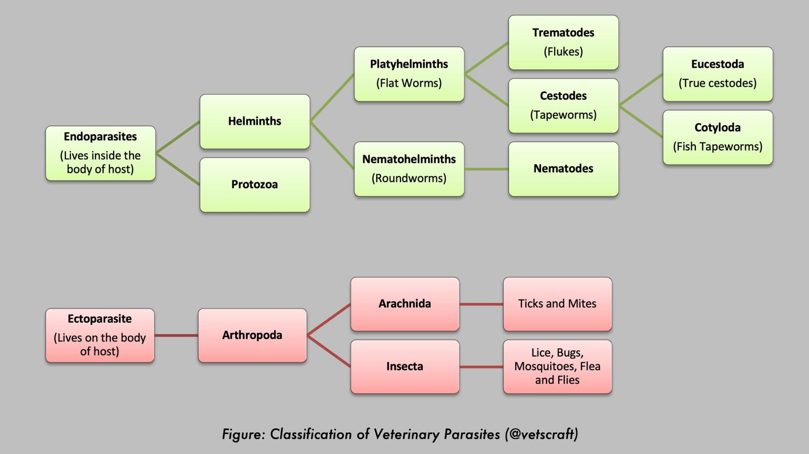
Prosthogonimus spp. are trematode parasites commonly known as oviduct flukes of poultry and wild birds. These parasites primarily inhabit the oviduct and Bursa of Fabricius, causing reproductive disorders, reduced egg production, abnormal eggs, and, in severe cases, peritonitis.
The life cycle involves aquatic snails and dragonfly nymphs as intermediate hosts. This article discusses the morphology, life cycle, pathogenesis, clinical signs, diagnosis, treatment, and prevention of Prosthogonimus infections in poultry.
Prosthogonimidae Family
Three oviduct flukes belonging to the family Prosthogonimidae are commonly studied:
- Prosthogonimus pellucidus or P. cuneatus
- Prosthogonimus macrorchis
- Prosthogonimus ovatus
1. Prosthogonimus pellucidus or P. cuneatus
- Family: Prosthogonimidae (Oviduct Flukes)
- Hosts: Chickens, turkeys, ducks, geese, and other domestic and wild birds
- Location: Bursa of Fabricius, oviduct, and posterior intestine
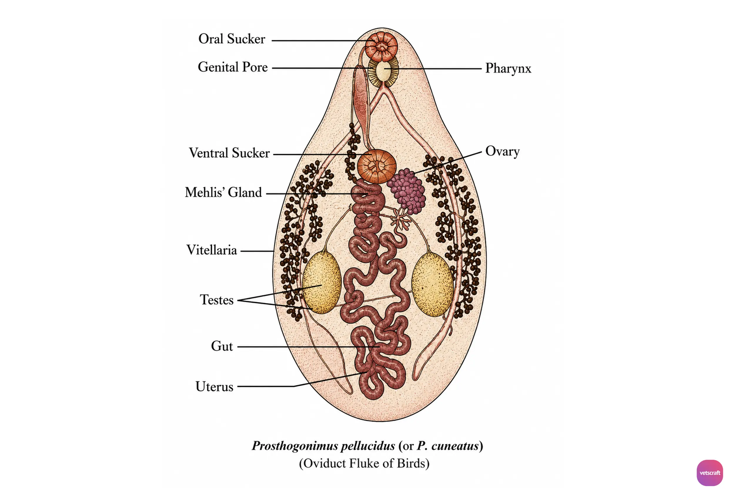
Morphological characteristics of Prosthogonimus pellucidus or P. cuneatus:
- Prosthogonimus pellucidus or P. cuneatus is a small fluke measuring 8–9 × 4–5 mm and is pale reddish-yellow in color when fresh.
- Pear-shaped, tapering anteriorly and broad or rounded posteriorly.
- The cuticle is covered with spines.
- The ventral sucker (VS) is located in the anterior half of the body.
- Irregularly oval testes lie horizontally in the middle of the body.
- The genital pore is situated adjacent to the oral sucker, and the cirrus sac is elongated, extending close to the oral sucker (OS).
- The ovary is highly lobulated and lies partly dorsal to or close to the ventral sucker.
- The vitellaria extend from the level of the ventral sucker to the posterior end of the testes and appear as grape-like clusters in the lateral fields.
- The eggs are operculated, dark brown in color, and bear a small spine at the pole opposite the operculum. They measure approximately 26–32 × 10–15 µm.
2. Prosthogonimus macrorchis
- Family: Prosthogonimidae (Oviduct Flukes)
- Hosts: Chickens, turkeys, ducks, geese, wild gallinaceous birds, and passerines (songbirds)
- Location: Bursa of Fabricius and oviduct
Prosthogonimus macrorchis is a small distome measuring 5–7 mm in length. The testes are relatively larger than those of P. pellucidus.
3. Prosthogonimus ovatus
- Family: Prosthogonimidae (Oviduct Flukes)
- Hosts: Gallinaceous birds and passerines (songbirds)
- Location: Bursa of Fabricius and oviduct
- Distribution: Europe, Africa, and Asia
Prosthogonimus ovatus is smaller than the two species described above, measuring 3–6 × 1–2 mm. Its testes are slightly elongated and lie side by side behind the mid-body region.
Life Cycle
Two intermediate hosts (IH) are required to complete the life cycle of Prosthogonimus spp.
- First IH (Aquatic Snails):
- P. pellucidus: Bithynia tentaculata
- P. macrorchis: Amnicola limosa
- P. ovatus: Bithynia leachi, Gyraulus albus
- Second IH:
- Dragonfly naiads (nymphs) – Tetragoneuria
The eggs hatch into miracidia, which enter aquatic snails. Sporocysts develop and produce cercariae. The redial stage is absent.
Cercariae are drawn into the anal openings of dragonfly naiads through the respiratory movements of these insects.
The cercarial tail is lost within the respiratory chamber of the nymph, and the metacercaria penetrates the musculature and encysts within the hemocoel.
Metacercariae may persist until the insect reaches maturity, and the definitive host (DH) becomes infected by ingesting either adult dragonflies or dragonfly nymphs (naiads).
Within the definitive host, the liberated immature flukes migrate to the cloaca and Bursa of Fabricius, where they mature. In adult birds, when the bursa has atrophied, the parasites migrate to the oviduct.
Pathogenesis
P. pellucidus is considered more pathogenic in chickens and turkeys than in ducks and geese.
A small number of flukes may not cause clinical disease. However, heavy infections can severely affect hens, causing marked irritation and significant impairment of egg production. Abnormal eggs may be produced, including soft-shelled eggs, shell-less eggs, or amorphous masses containing flukes. Albumen discharge from the cloaca may also occur. Most damage results from the flukes’ cuticular spines, which injure oviductal tissues and interfere with normal peristaltic activity.
The irritated oviduct readily undergoes retroperistaltic movements, resulting in rupture of yolks. Consequently, albumen and parasites may enter the peritoneal cavity and cause peritonitis, which can be fatal.
Secondary bacterial infections are not uncommon and may significantly worsen the condition.
Clinical Signs
- The disease is most commonly observed during spring and early summer.
- Initially, the general health of affected birds is not noticeably impaired.
- Birds show a marked tendency to remain in the nest. They become listless, develop a pendulous abdomen, and walk with their legs held widely apart.
- The irritated oviduct passes eggs rapidly, resulting in soft-shelled or shell-less eggs. Although the secretory glands function normally, their secretions are discharged separately.
- Egg laying is suspended, and affected birds become visibly ill.
- A milky discharge from the cloaca, consisting of lime deposits, may glue feathers around the vent. Feathers surrounding the cloaca become soiled with albumen and may contain yellowish-white strands and parasites.
- If peritonitis develops, the comb and wattles become cyanotic, and birds become prostrate and may die.
- If aseptic peritonitis develops, yolk material within the peritoneal cavity becomes inspissated and may obstruct intestinal motility.
- Adult flukes may occasionally be found within eggs.
Postmortem Lesions
- The oviduct shows varying degrees of catarrhal to croupous inflammation, with dirty caseous material in the lumen containing broken yolks and albumen.
- The parasites are not easily observed within the mucosal lining.
- In cases of peritonitis, the abdominal cavity contains turbid fluid, and organs may be adhered together by a cheesy exudate.
- Inspissated yolk material may be present between intestinal loops and around the oviduct.
- The serosal membranes exhibit marked congestion and hemorrhages.
Diagnosis
- Parasite eggs can be detected in cloacal discharges.
- In some cases, the parasites may have disappeared while the disease process persists. In such cases, eggs may still be found within the abdominal cavity during necropsy.
Treatment
- Albendazole: 10–50 mg/kg body weight.
- Praziquantel: 5–10 mg/kg body weight.
Prevention and Control
- Control aquatic snail intermediate hosts.
- Prevent birds from consuming dragonfly naiads in humid environments where these insects develop.