TABLE OF CONTENTS
Paragonimus westermani (Oriental Lung Fluke) and P. kellicotti: Morphology, Life Cycle, Pathogenesis, Diagnosis & Treatment
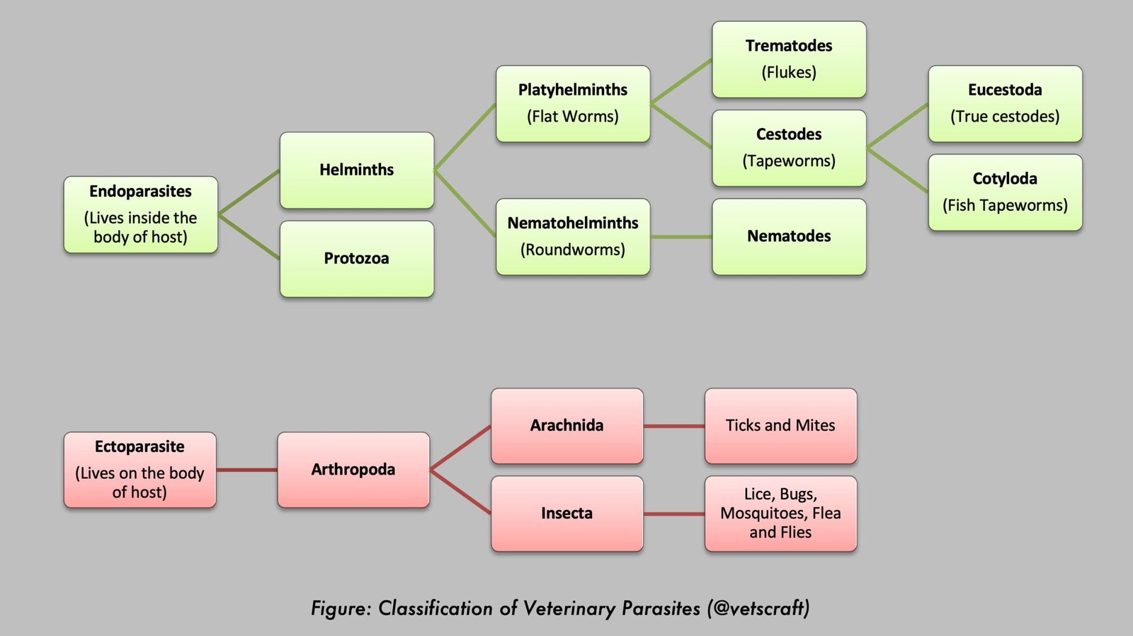
Paragonimus spp. are trematode parasites commonly known as lung flukes. These parasites infect a variety of mammals, including dogs, cats, pigs, wild carnivores, and humans. The most important species are Paragonimus westermani (Oriental Lung Fluke) and Paragonimus kellicotti (North American Lung Fluke). Adult flukes primarily inhabit the lungs, causing respiratory disease and occasionally affecting other organs such as the brain and muscles.
Paragonimus westermani (Oriental Lung Fluke)
- Family: Paragonimidae (Lung Flukes)
- Hosts: Pigs, dogs, cats, foxes, goats, cattle, and humans
- Location: Lungs and, less commonly, the brain, spinal cord, and striated muscles
- Distribution: China, Southeast Asia, and Far Eastern countries
Paragonimus kellicotti (North American Lung Fluke)
- Family: Paragonimidae (Lung Flukes)
- Hosts: Wild animals, cats, dogs, and pigs. The primary host is probably the mink, and the muskrat may also serve as a natural host.
- Location: Lungs
- Distribution: North America
Morphology
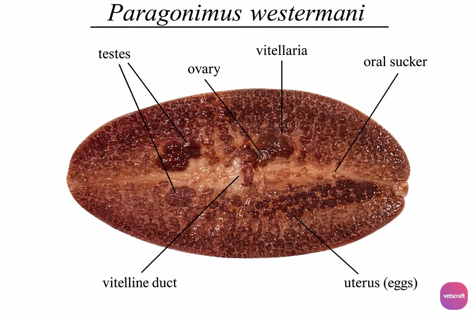
- Paragonimus spp. are ovoid, plump distomes, and the adults live in pairs within cysts in the lungs.
- They are reddish-brown in color and measure 7.5–16 × 4–8 mm.
- The tegument is covered with spines. The spines of P. westermani are large with bifid tips, whereas those of P. kellicotti are larger and possess multiple points.
- The oral sucker (OS) is ventro-terminal. The ventral sucker (VS) is situated slightly anterior to the middle of the body.
- The genital pore is located immediately behind the VS.
- The testes are branched and lie side by side below the VS in the posterior half of the body.
- The ovary is pretesticular and positioned just below the VS.
- The uterus forms a thick coil and is located to the right of the midline.
- The eggs are yellowish-brown and measure 75–118 × 42–67 µm. They are operculated, and the shell is thickened at the pole opposite the operculum.
Life Cycle
The eggs are laid within the cysts and escape through connecting channels into the bronchi or when the cysts rupture. They pass up from the lungs and may be found in the sputum, which has a characteristic rusty color. Animals swallow the mucus; therefore, the eggs are also found in the feces.
After development for 2–7 weeks (16 days under optimal conditions), the miracidium hatches and penetrates the first intermediate host (aquatic or amphibious snails of the genera Melania, Ampularia, and Semisulcospira), where it develops into sporocysts, rediae, and cercariae over a period of 78–93 days. The cercaria has an oval body and a short tail (microcercous cercaria) and is released from the snail, swimming in water until it encounters a suitable second intermediate host.
The cercariae penetrate the second intermediate host, which is a crustacean such as a crab or crayfish (Astacus, Cambarus), and encyst. The metacercariae are found in the heart, liver, and muscles and mature over six to seven weeks.
The definitive hosts, including dogs, cats, pigs, and humans, become infected by consuming infected crustaceans. Metacercariae released from injured or degenerating crustaceans can survive in water for up to three weeks and may be ingested by the definitive host.
The metacercariae excyst in the intestine, and the young flukes penetrate the intestinal wall and migrate through the peritoneal cavity for 1–14 days. They then pass through the diaphragm and pleural cavity before entering the lungs approximately 5–23 days after infection. They may also migrate to other organs from this location.
The parasite penetrates the pulmonary parenchyma and forms a cystic cavity in which it matures to the adult stage. Virtually all growth occurs within the pulmonary cyst. Communication with the bronchioles is established within five weeks, and the prepatent period is 30–36 days.
The cyst usually contains two parasites surrounded by purulent fluid mixed with blood and eggs. The interior surface of the cyst is partially epithelialized by cells derived from the bronchioles. In cats, 1,000–2,000 eggs may be laid per fluke per day.
The worms may also reach other organs and tissues, such as the brain and striated muscles. However, when this occurs, completion of the life cycle is not achieved because the eggs deposited cannot exit these sites. The period from infection to oviposition is 65–90 days. Infections may persist for up to 20 years in humans.
Pathogenesis
Migrating immature flukes cause eosinophilic peritonitis, pleuritis, myositis, and multifocal hemorrhage.
In infected animals, chronic bronchiolitis, hyperplasia of the bronchiolar epithelium, and chronic eosinophilic granulomatous pneumonia develop, associated with degenerating ova within the alveolar tissue.
Clinical Signs
Infected animals are lethargic. The acute phase (invasion and migration) may be characterized by diarrhea, abdominal pain, fever, intermittent cough, weight loss, urticaria, hepatosplenomegaly, pulmonary abnormalities, and marked eosinophilia.
During the chronic phase, pulmonary manifestations include cough, expectoration of discolored sputum, hemoptysis, and radiographic abnormalities of the chest. Dyspnea associated with pneumothorax has been reported in cats.
Extrapulmonary adult parasites lodged within brain cysts may cause neurological disorders.
Pleural effusion, a condition characterized by excessive fluid accumulation around the lungs, is another clinical sign of infection.
Diagnosis
- Pulmonary cases are readily diagnosed by detecting eggs in the sputum or feces.
- Lesions may be detected radiographically (X-ray) as early as 3–4 weeks after infection. The lesions are most commonly found in the right caudal lung lobe.
- Diagnosis of cysts in other parts of the body is extremely difficult; however, various serological tests such as CFT and ELISA may be employed.
Treatment
- Albendazole: 50–100 mg/kg/day for 14–21 days. This reduces egg shedding within 8 days, causes morphological degeneration of adult flukes, and reduces pulmonary lesions in cats.
- Fenbendazole: 50 or 100 mg/kg/day in two divided doses for 10–14 days.
- Bithionol: 100 mg/kg/day for 7 days.
- Niclofolan: 1 mg/kg/day for 3 days or two doses of 2 mg/kg administered on alternate days.
Prophylaxis
- Freshwater crustaceans (crabs and crayfish) should not be consumed raw.
- Avoid drinking untreated water from endemic areas.
- Control snail intermediate hosts.
- Treat infected animals and humans.
- Ensure hygienic disposal of animal feces and human waste.