TABLE OF CONTENTS
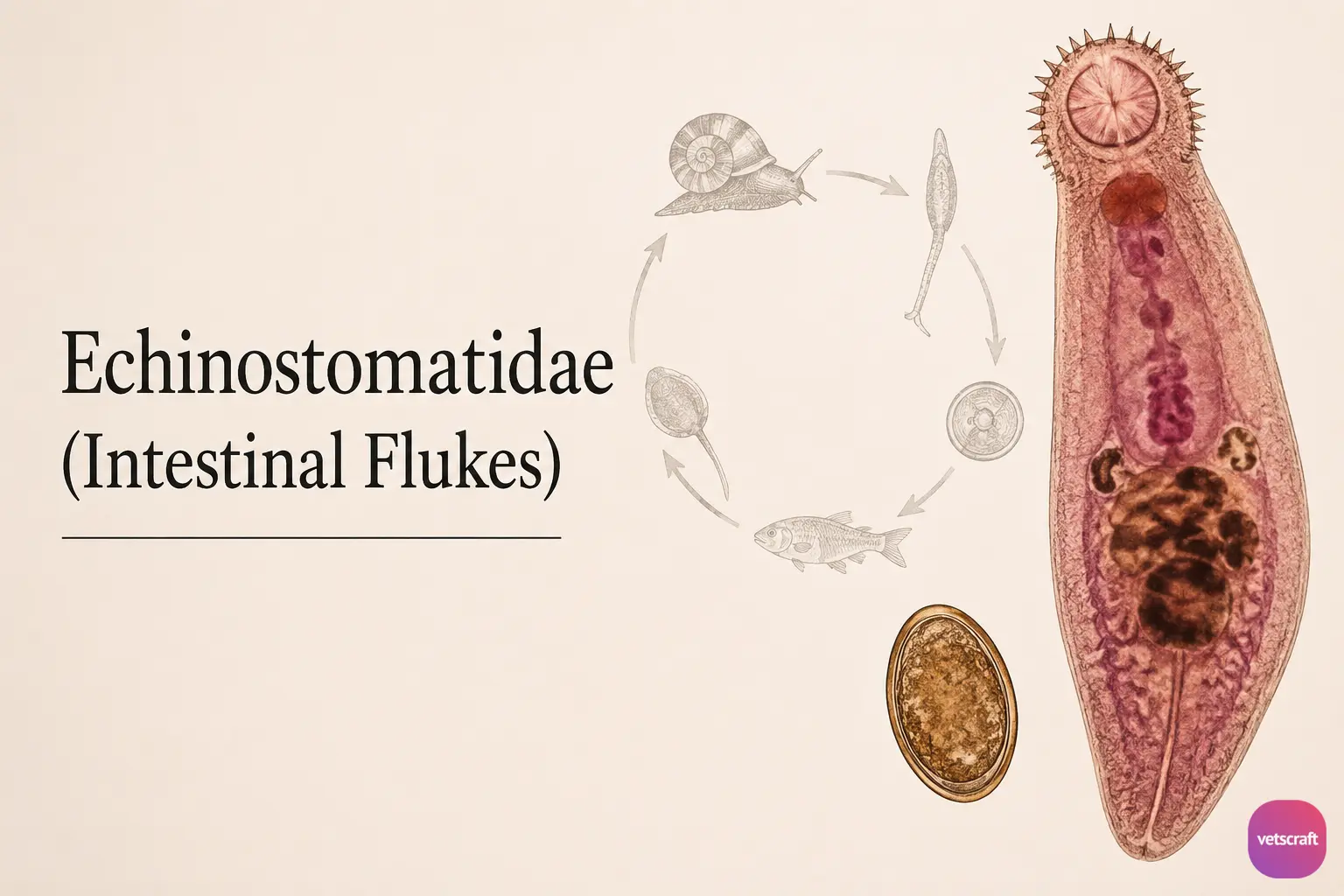
Echinostomatidae (Intestinal Flukes): Morphology, Life Cycle, Hosts, Diagnosis & Treatment
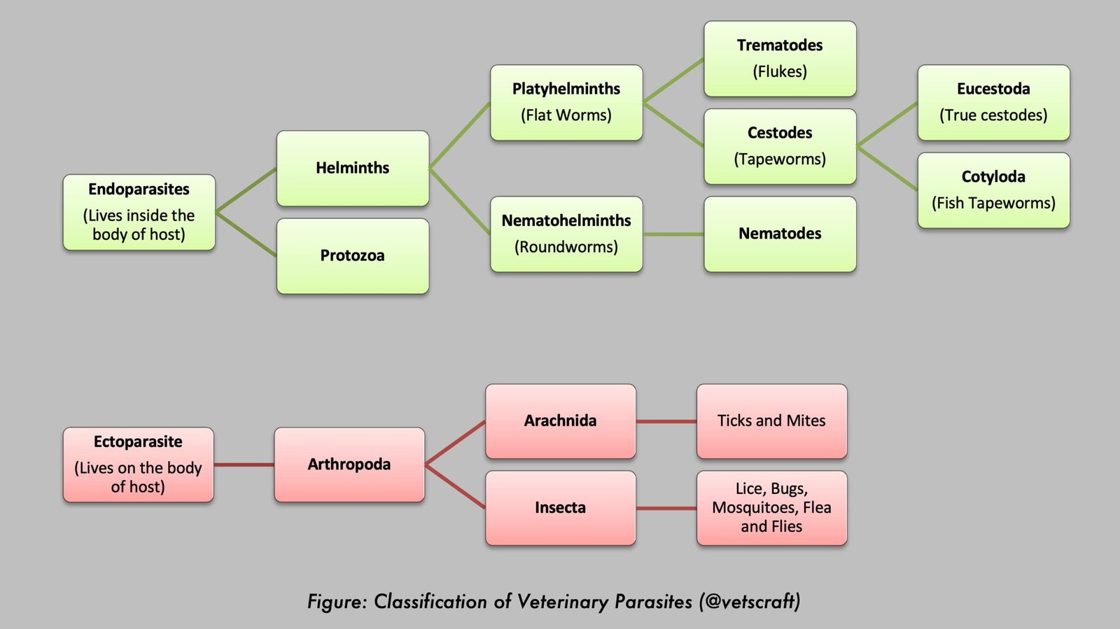
Echinostomatidae are intestinal trematodes (flukes) that infect birds, mammals, and humans worldwide. Members of the genus Echinostoma are characterized by a distinctive collar of spines surrounding the oral sucker and require one or more intermediate hosts to complete their indirect life cycle. This article describes the morphology, hosts, life cycle, clinical signs, diagnosis, treatment, and prevention of echinostome infections.
- Family: Echinostomatidae (intestinal flukes)
- Genus: Echinostoma (E. revolutum sensu lato, E. paraulum, E. ilocanum, E. hortense)
Morphology
- Flukes are elongate, much longer than wide, and measure approximately 2–10 mm in length and 1–2 mm in width.
- They are spiny-collared intestinal flukes. The distinctive head collar bears numerous spines (arrows) and surrounds the oral sucker/aperture.
- The oral sucker (OS) is smaller, while the ventral sucker (VS) is more prominent. The OS and VS are located relatively close to one another.
- The oral sucker is surrounded by a head collar with spines. The number of spines varies among species, but there are usually between 37 and 51. These spines are arranged in one or two rows around the sucker, and their arrangement may be a characteristic feature of an Echinostoma species.
- E. revolutum – The OS bears 37 spines, with five corner spines on each side.
- E. paraulum – The OS bears 37 spines, of which 27 are arranged in double rows, with five corner spines on each side.
- The digestive system consists of a pharynx, esophagus, and an excretory pore. The intestine extends to the posterior extremity.
- The testes are entire or lobed, arranged in tandem or slightly diagonal, and situated in the posterior half of the body. A single ovary is located near the large paired testes. The vitellaria consist of coarse follicles occupying the lateral fields.
Hosts
Echinostomatidae utilize a complex indirect life cycle involving definitive and intermediate hosts. Adult flukes inhabit the intestine of the definitive host, while larval development occurs within one or more intermediate hosts before transmission to the definitive host.
| Species | Host and Location | First IH | Second IH |
|---|---|---|---|
| E. revolutum | Ducks, geese, birds, humans – rectum, ceca | Stagnicola, Helisoma, Planorbis, Physa, Lymnaea | Vivipara, Fossaria, tadpoles of Rana |
| E. paraulum | Ducks, pigeons, humans – intestine | Unknown | Unknown |
| E. ilocanum | Humans, rats, dogs – intestine | Gyraulus | Vivipara, Fossaria |
| E. hortense | Humans, rats, dogs – intestine | Snail | Frog, loach |
Definitive Host (DH)
Birds, partridges, rats, dogs, and humans serve as definitive hosts for Echinostomatidae (intestinal flukes).
Intermediate Host (IH)
The first intermediate host is always a snail (Families: Planorbidae, Lymnaeidae, and Bulinidae). In some cases, the same first intermediate host may also act as the second intermediate host. Depending on the species, several animals may serve as the second intermediate host, including other snails, bivalves, fish, salamanders, and tadpoles.
Life Cycle
Like many trematodes, echinostomid flukes undergo a multi-host (indirect) life cycle. Unembryonated eggs are passed in the feces of infected definitive hosts and develop in water. Miracidia usually take about 3 weeks to mature before hatching, after which they swim freely and penetrate the first intermediate host, a snail.
The intramolluscan stages include a sporocyst stage, one or two generations of rediae, and cercariae, which are released from the snail. The cercariae may encyst as metacercariae within the same first intermediate host or leave the host and penetrate a new second intermediate host. The definitive host becomes infected after ingesting metacercariae within an infected second intermediate host.
Metacercariae excyst in the duodenum, and adults reside in the small intestine (for some species, occasionally in the bile ducts or large intestine).
Clinical Symptoms
- Catarrhal inflammation often occurs due to the penetration of the sharp spined collar into the intestinal mucosa, creating ulcerative lesions.
- In heavy infections, abdominal pain, fatigue, and weight loss may occur.
- Peripheral eosinophilia is usually present.
- A small number of fatal cases of Artyfechinostomum (Echinostoma) malayanum, in which heavy worm burdens caused anemia, malnutrition, or intestinal perforation, have been reported.
Diagnosis
Diagnosis is based on the microscopic identification of yellow-brown eggs in stool samples. Methods such as the Kato-Katz procedure can be used for detection. The Kato technique (also called the Kato-Katz technique) is a laboratory method used to prepare human stool samples prior to examination for parasite eggs.
Treatment
Oxyclozanide is commonly used at a dose rate of 15–30 mg/kg body weight. Rafoxanide may also be effective against certain echinostome infections.
Prophylaxis
- Implement snail control measures.
- Avoid consuming frogs, snails, and loaches.
- Echinostome cercariae have been used in the biological control of schistosomes, as their larval stages exhibit predatory activity against the larval stages of schistosomes.