
TABLE OF CONTENTS
Leucocytozoon smithi (Akiba smithi): Life Cycle, Pathogenesis, Diagnosis & Treatment
Leucocytozoon smithi (Akiba smithi) is a hemoprotozoan parasite of domestic and wild turkeys that belongs to the family Leucocytozoidae. It is transmitted by black flies (Simulium spp.) and is an important cause of leucocytozoonosis in turkeys, particularly affecting young birds. Severe infections may result in high mortality and significant economic losses in turkey production.
This article provides concise veterinary notes on Leucocytozoon smithi (Akiba smithi), including its taxonomy, morphology, life cycle, pathogenesis, clinical signs, postmortem lesions, diagnosis, and treatment.
Parasite Overview
- Host: Domestic and wild turkeys
- Vector: Simulium spp.
- Common Name: Turkey leucocytozoon
Taxonomical Classification
- Domain: Eukaryota
- Kingdom: Protista
- Phylum: Apicomplexa
- Class: Sporozoea
- Order: Haemosporida
- Family: Leucocytozoidae
- Genus: Leucocytozoon (Akiba)
- Species: Leucocytozoon smithi (Akiba smithi)
Morphology
Leucocytozoon smithi (Akiba smithi) resembles L. simondi. The mature gamonts are elongate, and the host cell is greatly distorted and elongated. The nucleus of the host cell is elongated, forming a dark band on one side of the parasite, and it is frequently split to form a dark band on each side of the parasite.
Life Cycle
The life cycle is similar to that of L. simondi, but megaloschizonts do not occur. The prepatent period is 9 days.
Pathogenesis
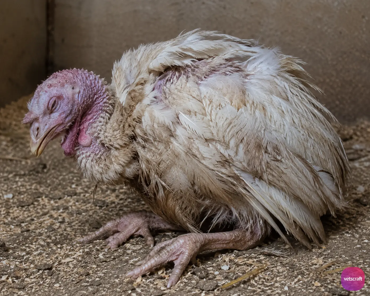
Leucocytozoon smithi (Akiba smithi) may be extremely pathogenic in young turkeys, with mortality reaching up to 90%. Clinical signs include anorexia, emaciation, debility, leg weakness, and incoordination during the later stages of the disease.
Birds usually die within 2–3 days after the onset of clinical signs. However, some birds may develop a chronic form of the infection, characterized by coughing and moist bronchitis.
Post Mortem Lesions
Jaundice, congestion of the duodenum, splenomegaly, and congestion of the lungs and kidneys.
Diagnosis
Examination of blood smears.
Treatment
Sulphaquinoxaline may be tried.

