
TABLE OF CONTENTS
Leucocytozoon simondi (Akiba simondi): Morphology, Life Cycle, Pathogenesis & Treatment
Leucocytozoon simondi (Akiba simondi) is a hemoprotozoan parasite of domestic and wild ducks belonging to the family Leucocytozoidae. It is transmitted by the bite of Simulium blackflies and occasionally Culicoides biting midges. The parasite undergoes schizogony in various tissues and gametogony within blood cells, resulting in significant disease, particularly in young ducks. This article covers the taxonomy, morphology, life cycle, pathogenesis, diagnosis, treatment, and control of Leucocytozoon simondi for veterinary students and professionals.
Parasite Overview
- Host: Domestic and wild ducks
- Vector: Simulium spp.
- Predilection Site: Schizogony occurs in the endothelial and parenchymal cells of the liver, kidneys, lungs, and intestinal lymphoid cells. Gametogony occurs in the erythrocytes and lymphocytes.
Taxonomical Classification
- Kingdom: Protista
- Phylum: Apicomplexa
- Class: Sporozoea
- Order: Haemosporida
- Family: Leucocytozoidae
- Genus: Leucocytozoon (Akiba)
- Species: Leucocytozoon simondi (Akiba simondi)
- Common Name: Duck leucocytozoon
Morphology
The gamonts of Leucocytozoon simondi (Akiba simondi) are elongate or ovoid in shape. The infected host cells become distorted and spindle-shaped.
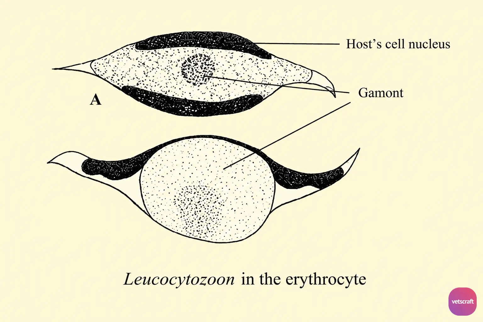
The host cell nucleus is also elongated and forms a long, thin, dark, crescent-shaped band along one side of the parasitized cell. In some species, the host cell nucleus is split to form a dark band on either side of the parasite.
In the macrogametes, the nucleus is compact, and several vacuoles may occur in the cytoplasm, whereas in the microgametes, the nucleus is diffuse and stains pale blue.
Transmission and Life Cycle
Sporozoites are introduced when Simulium or Culicoides flies suck blood from birds. Following inoculation, the sporozoites are carried by the blood to various organs. The first generation of schizogony takes place in the Kupffer cells of the liver, where small schizonts produce merozoites.
Some of the first-generation merozoites enter blood cells to become gamonts, while others initiate hepatic schizonts and megaloschizonts. The hepatic schizonts occur in the liver parenchymal cells and produce a number of cytomeres. The cytomeres then undergo multiple fission to form a large number of small merozoites.
The megaloschizonts are large and more numerous than the hepatic forms. They occur in lymphoid cells or macrophages (brain, liver, lungs, kidneys, intestinal tissues, and lymphoid tissues) 4–6 days after infection. They contain a large number of cytomeres, which, in turn, produce numerous merozoites.
Following rupture of the hepatic schizonts and megaloschizonts, merozoites are released into the blood and then enter erythrocytes, where they become gamonts 6–7 days after infection. Development in the vector is similar to that of Plasmodium in mosquitoes.
Pathogenesis
Leucocytozoon simondi is highly pathogenic in young ducks. The clinical signs of the disease include listlessness, anorexia, and rapid breathing due to the presence of a large number of megaloschizonts in the pulmonary capillaries. Death may occur within a day or so, and affected birds may exhibit nervous disorders before death.
The disease in older birds is less acute and develops more slowly. Affected birds become emaciated and listless but seldom die in less than four days after the onset of the disease.
Diagnosis
Demonstration of gamonts in blood smears and megaloschizonts in impression smears from the lungs.
Treatment
No effective treatment is available.
Control
- Control Simulium flies.
- Young ducklings should be isolated from older birds, as the latter may serve as carriers.

