
TABLE OF CONTENTS
Haemoproteus columbae (Pigeon Malaria-Like Parasite): Morphology, Life Cycle, Pathogenesis, Diagnosis & Treatment
Haemoproteus columbae, commonly known as the pigeon malaria-like parasite, is a hemoprotozoan parasite of domestic and wild pigeons. It belongs to the phylum Apicomplexa and is transmitted by the pigeon louse fly, Pseudolynchia canariensis. The parasite undergoes schizogony in the endothelial cells of blood vessels and gametogony in erythrocytes, while sexual reproduction occurs in the insect vector.
Although infections are generally mild, H. columbae may occasionally cause anemia, emaciation, and reduced performance in pigeons. This article provides a detailed overview of its taxonomy, morphology, life cycle, pathogenesis, diagnosis, postmortem lesions, and treatment.
Parasite Overview
- Host: Domestic and wild pigeons
- Vector: Pseudolynchia canariensis
- Predilection Site: Schizogony occurs in the endothelial cells of blood vessels, and gametogony (gamonts) occurs in the erythrocytes (sexual reproduction occurs in the vector).
Taxonomical Classification
- Kingdom: Protista
- Phylum: Apicomplexa
- Class: Sporozoea
- Order: Haemosporida
- Family: Haemoproteidae
- Genus: Haemoproteus
- Species: Haemoproteus columbae
- Common Name: Pigeon haemoproteid (pigeon malaria-like parasite)
Morphology
Haemoproteus columbae also forms pigment granules (haemozoin) from the host cell hemoglobin. Gamonts are found in the erythrocytes. They are elongated and crescent-shaped, encircling the nucleus of the host cell and giving a halter-shaped appearance.
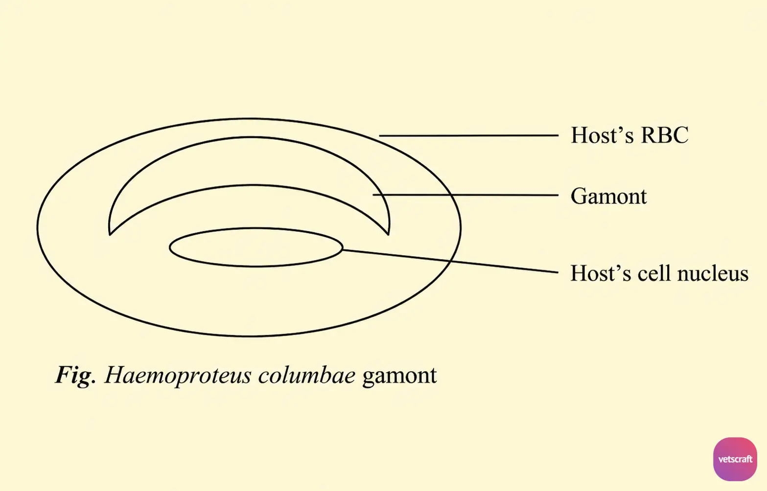
The nucleus of the host cell may be displaced. In the macrogamonts, the nucleus is compact, and pigment granules are dispersed throughout the cytoplasm, whereas in the microgamonts, the nucleus is diffuse, and the pigment granules are collected into a spherical mass.
Transmission and Life Cycle
Development in the Pigeon
When an infected hippoboscid fly bites a pigeon, sporozoites are inoculated. The sporozoites enter the bloodstream and penetrate the endothelial cells of blood vessels, where they develop into early schizonts.
The early schizonts have minute cytoplasmic bodies and a single nucleus, but as a result of repeated nuclear divisions, 15 or more uninucleate cytomeres are produced.
The nuclei of the cytomeres then undergo repeated division until the endothelial cell is filled with a large number of multinucleate cytomeres surrounded by a fine cyst wall.
Each cytomere produces an enormous number of minute merozoites. Due to excessive pressure, the infected endothelial cell may rupture, releasing the cytomeres.
These cytomeres accumulate in the capillaries, causing blockage, but they soon rupture, and the merozoites escape into the bloodstream. The merozoites then enter red blood cells and develop into gamonts.
Development in the Vector
It is similar to that of Plasmodium in the mosquito. The motile zygote (ookinete) migrates to the outer surface of the midgut, where sporogony takes place to produce a large number of sporozoites.
These sporozoites are released into the body cavity of the insect and then pass to the salivary glands. They are injected into a new host when an infected fly feeds.
Pathogenesis
H. columbae is generally less pathogenic, but it may occasionally cause anemia and emaciation in adult pigeons.
Postmortem Lesions
The liver and spleen are enlarged and dark in color.
Diagnosis
- Demonstration of gamonts in blood smears.
- Demonstration of schizonts in impression smears from the lungs.
Treatment
- Quinacrine is effective against young gamonts.

