
TABLE OF CONTENTS
Equine Babesiosis (Equine Piroplasmosis): Causes, Diagnosis & Treatment
Equine babesiosis, also known as equine piroplasmosis, is a tick-borne hemoprotozoan disease of horses, donkeys, mules, and zebras caused by Babesia caballi and Babesia equi (currently classified as Theileria equi). The disease is characterized by fever, anemia, jaundice, lethargy, and reduced athletic performance, resulting in significant health and economic impacts on equine populations worldwide.
Transmission occurs primarily through infected hard ticks, although iatrogenic transmission through contaminated blood or instruments may also occur. Diagnosis is based on clinical signs, microscopic examination of blood smears, and serological or molecular tests such as IFAT and PCR. Effective management relies on early diagnosis, appropriate antiprotozoal therapy, supportive care, and tick control measures.
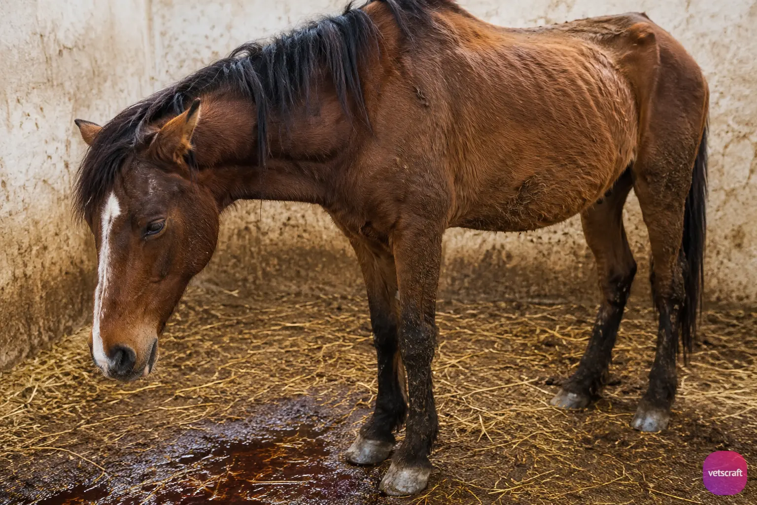
Species of Babesia affecting horses, mules, donkeys, and zebras include Babesia caballi and Babesia equi.
Babesia equi (Theileria equi)
Morphology
- Merozoites are small (2 µm), round, amoeboid, or pyriform in shape.
- The pyriform type divides into four daughter organisms (arranged in tetrads), forming a Maltese cross.
- There may be up to eight merozoites in a single RBC.
Life Cycle
Refer to the general life cycle of Babesia species.
Vector
Dermacentor reticulatus, Hyalomma excavatum, H. dromedarii, H. marginatum, R. bursa, R. evertsi, and H. anatolicum anatolicum.
The mode of transmission is transovarial in H. anatolicum, whereas it is transstadial in the other tick species. Exoerythrocytic schizogony in lymphocytes has been reported.
Pathogenesis and Clinical Signs
Babesia equi is more pathogenic than Babesia caballi. The incubation period is 8–10 days. The first clinical sign is an increase in body temperature (41–42°C), followed by anemia and marked hemoglobinuria, which is not observed in B. caballi infections.
Affected animals show listlessness, depression, loss of appetite, marked thirst, watery eyes, and swollen eyelids. Hemorrhages are present on the mucous membranes of the nasal passages, vagina, and third eyelids.
Edema develops in the dependent parts of the body, especially the ventral aspect, legs, and head. The feces are hard and covered with yellow mucus. In peracute cases, death occurs 1–2 days after the onset of clinical signs.
Postmortem Lesions
- Accumulation of fluid in the pericardial sac and body cavity
- Jaundice
- Petechial hemorrhages in the mucous membranes of the stomach and intestines
- Enlargement of the liver and spleen
- Flabby kidneys
- Pulmonary edema
Immunity
- Recovered animals are immune to reinfection.
- There is no cross-immunity between the two species.
Diagnosis
- Clinical signs.
- Examination of a peripheral blood smear.
- IFAT is the most commonly used test.
Treatment
- Amicarbalide: 9 mg/kg SC or IM.
- Imidocarb: 2.2 mg/kg IM, administered four times at 3-day intervals; however, it is toxic in donkeys.
- Berenil: 11 mg/kg IM.
Babesia caballi
Morphology
Merozoites are large, resembling those of B. bigemina, and are round, oval, or pyriform in shape.
Life Cycle
Babesia caballi is transmitted through transovarial or transstadial transmission by Hyalomma anatolicum and H. marginatum.
Pathogenesis and Clinical Signs
The pathogenesis varies considerably among animals. The disease may follow either an acute or chronic course. B. caballi infection is characterized by fever, icterus, and severe anemia without hemoglobinuria. Affected animals may also develop gastroenteritis and posterior paralysis.
Diagnosis
Examination of a blood smear.
Treatment
- Amicarbalide: 22 mg/kg body weight IM
- Imidocarb: 4 mg/kg body weight
Conclusion
Equine babesiosis (equine piroplasmosis) is an important tick-borne disease affecting equids worldwide. Early diagnosis, prompt treatment, and effective tick control are essential for reducing disease severity and preventing transmission. Understanding the morphology, life cycle, clinical signs, diagnosis, and treatment of Theileria equi and Babesia caballi is important for veterinary students, clinicians, and equine practitioners.

