
TABLE OF CONTENTS
Babesia Species: Life Cycle, Pathogenesis, Transmission & Veterinary Importance
Babesia species are tick-borne, intraerythrocytic protozoan parasites of major veterinary importance, causing babesiosis in a wide range of domestic and wild animals. These hemoprotozoa invade erythrocytes, resulting in anemia, fever, hemoglobinuria, and, in severe cases, multi-organ dysfunction. The life cycle of Babesia is heteroxenous, involving both a vertebrate host and a tick vector, with transmission occurring through transovarian or transtadial routes depending on the species.
Merozoites of Babesia species are found in erythrocytes and usually lie in pairs at an angle with their narrow ends opposed. They are relatively large and may be pyriform, round, or oval. When stained with Giemsa or Leishman stain, the cytoplasm of the merozoites stains blue, and the chromatin mass located at one pole stains red.
A strand of chromatin may extend from the chromatin mass. In erythrocytes, they multiply by binary fission, endodyogeny, endopolygeny, or schizogony.
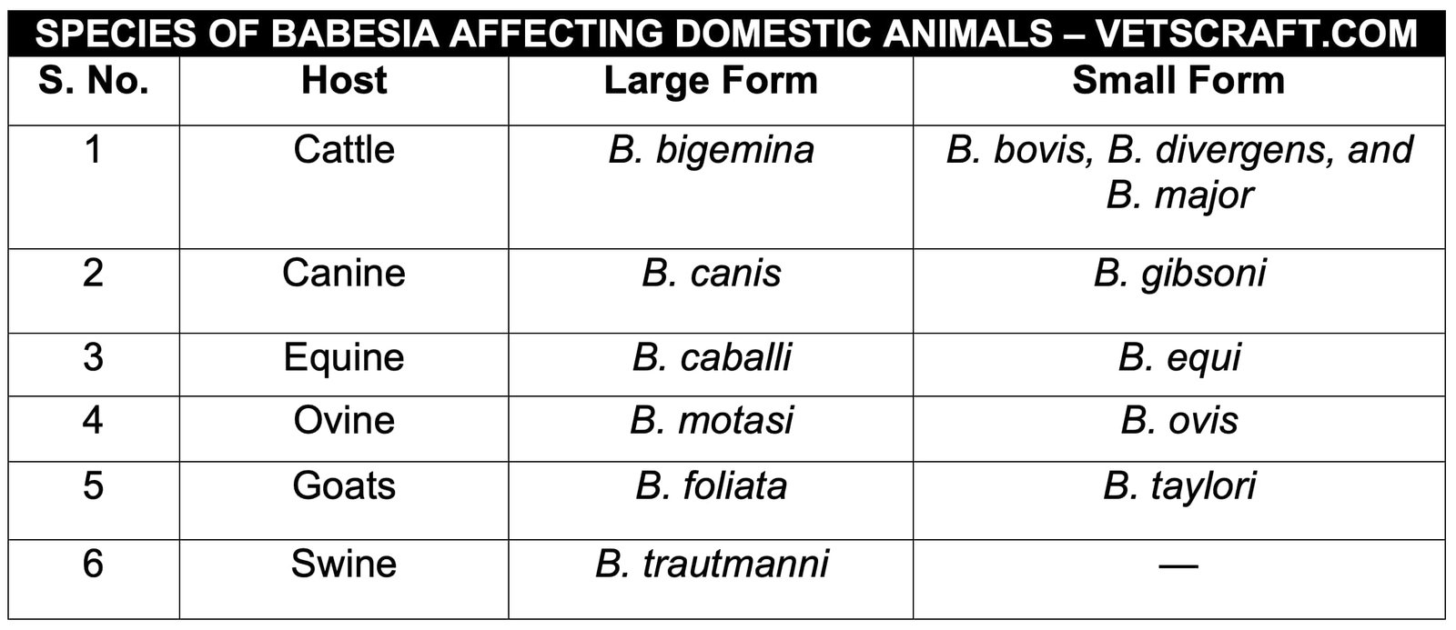
Based on the size and position of the merozoites in erythrocytes, the species within this genus are classified as larger forms (3 µm) and smaller forms (less than 2.5 µm). The larger forms are more pathogenic than the smaller forms. In erythrocytes, the merozoites of the larger forms lie at an acute angle, whereas those of the smaller forms lie at an obtuse angle.
The species of Babesia affecting domestic animals:
General Life Cycle Pattern of Babesia
Babesia is a heteroxenous parasite that requires two hosts. Its asexual reproduction occurs in the vertebrate host, whereas sexual reproduction occurs in the tick vector. If a one-host tick (Boophilus spp.) acts as the vector, Babesia organisms are transmitted from one generation of ticks to the next through the ova. This is known as transovarian transmission and occurs in B. bigemina.
In this mode of transmission, the adult female tick acquires the infection while feeding on an infected animal. After further development within the adult tick, the organisms are transmitted to the next generation of larvae through the ova. These newly hatched larvae are then capable of transmitting the infection to a susceptible host.
Whereas, if a two-host or three-host tick acts as the vector, transmission of Babesia organisms from one developmental stage to the next is known as transtadial transmission, which occurs in B. canis in dogs. In this case, either the larval or nymphal stage of the tick acquires the infection while feeding on an infected animal, and the ingested organisms undergo further development within the tick.
By the time the tick molts to the next stage (e.g., larva to nymph or nymph to adult), the newly emerged nymph or adult is capable of transmitting the infection to a susceptible host.
The infection is transmitted from one vertebrate host to another through tick bites. Adult ticks and nymphs usually transmit the larger forms of Babesia, whereas the smaller forms are transmitted by larvae and adults.
Development in the Vertebrate Host
When an infected tick feeds on a susceptible animal, sporozoites are introduced into the host through the saliva. These sporozoites invade erythrocytes, where they multiply by binary fission, endodyogeny, endopolygeny, or schizogony to produce merozoites.
In some species, two merozoites are formed, whereas in others, four merozoites may develop within a single erythrocyte. The merozoites are released by mechanically rupturing the erythrocyte and subsequently invade new erythrocytes. This asexual phase continues indefinitely.
Finally, two types of merozoites are produced, namely paired or single pyriform forms and ovoid forms. The latter are considered the precursors of the sexual phase. Both types are found in the peripheral blood. Further development occurs in the tick after it feeds on an infected animal.
Development in the Invertebrate Host (Tick Vector)
Transovarian Transmission
When an adult female tick feeds on an infected animal, erythrocytes containing merozoites are ingested. In the tick gut, the merozoites are released following lysis of the erythrocytes. Of the two forms of merozoites, the pyriform forms are destroyed, whereas the ovoid forms enter the gut epithelial cells, where they develop into bizarre-shaped stages with numerous protrusions called Ray’s bodies or Strahlenkörper bodies.
Initially, they are tetranucleate bodies, but subsequently they give rise to uninucleate bodies through division. Two uninucleate Ray’s bodies then fuse to produce a zygote. Each zygote, in turn, produces a single kinete containing an internal vacuole.
These motile kinetes leave the intestinal cells and enter the hemolymph, where they invade cells of various organs, including the Malpighian tubules, muscle fibers, hemocytes, and developing ova of the tick. In these organs, the kinetes become non-motile and develop into polymorphic forms.
These polymorphic forms subsequently produce numerous uninucleate cytomeres, which in turn produce fission bodies, followed by sporokinetes (vermicules). The infection is then transmitted to the next generation when the larvae hatch (transovarian transmission). The sporokinetes subsequently migrate to the hemolymph and salivary glands, where they develop into large multinucleate sporonts (YS & ES) that give rise to thousands of sporozoites.
Transstadial Transmission (Stage-to-Stage Transmission)
This type of transmission occurs in the life cycle of B. canis in Rhipicephalus sanguineus. If the larval stage acquires the infection from an infected dog, the nymphal stage will be capable of transmitting the infection to a susceptible host. Similarly, if the nymphal stage acquires the infection, the adult stage will transmit the infection.
When larvae or nymphs feed on an infected animal, merozoites are ingested with the blood meal. The erythrocytes containing merozoites undergo lysis in the tick gut, releasing the merozoites.
The released merozoites then migrate to phagocytes located in the hypodermis, where they multiply to produce pseudocysts within 7 days. By 11–15 days of tick feeding, club-shaped stages are formed within the pseudocysts. These club-shaped stages are released and subsequently invade muscle cells, where they round up and divide repeatedly to produce large numbers of small ovoid forms.
When the tick molts to the next stage (larva to nymph or nymph to adult), the small ovoid forms migrate to the salivary glands, where they undergo binary fission to produce large numbers of sporozoites. When an infected tick feeds on a susceptible animal, these sporozoites are introduced into the host.
The inoculated sporozoites invade erythrocytes, where they multiply by multiple fission to produce merozoites.
General Pathogenesis of Babesiosis
- Release of pharmacologically active substances
- Anemia
- Glomerulonephritis
- CNS damage
1. Release of Pharmacologically Active Substances
- Activation of prekallikrein to kallikrein usually occurs 1–2 days before parasites appear in the blood. As a result, plasma kallikrein levels increase markedly by 3 days after infection.
- This elevated level of kallikrein increases vascular permeability and vasodilation, leading to circulatory stasis and shock.
- It also triggers intravascular coagulation.
2. Anemia
Anemia is associated with:
- Mechanical rupture of erythrocytes by emerging merozoites.
- Direct removal of non-infected RBCs by phagocytosis.
- Increased osmotic fragility of non-infected RBCs, which may predispose them to spontaneous lysis.
- Adsorption of Ag–Ab complexes onto the surface of RBCs, leading to their removal by phagocytosis.
3. Glomerulonephritis
Glomerulonephritis is associated with glomerular deposition of IgG and the third component of complement (C3).
4. CNS Damage
CNS damage is a characteristic feature of B. bovis and B. canis infections. In these infections, parasitized cells accumulate in the brain capillaries, leading to obstruction of blood flow.
In addition, the infected cells adhere to one another and to the vascular endothelium due to enzymatic activity. As a result, neurological signs develop.

