
TABLE OF CONTENTS
Bovine Babesiosis (Redwater Fever or Texas Fever): Clinical Signs, Diagnosis & Treatment
Bovine babesiosis, also known as Redwater Fever or Texas Fever, is an economically important tick-borne hemoprotozoan disease of cattle caused by Babesia species. The disease is characterized by fever, hemolytic anemia, hemoglobinuria, jaundice, and significant production losses, with severe cases resulting in death. The principal causative species include Babesia bigemina and Babesia bovis, both of which are transmitted primarily by hard ticks through transovarian and, in some species, transtadial transmission.
This article provides veterinary notes on bovine babesiosis, including the important Babesia species affecting cattle, morphology, life cycle, pathogenesis, clinical signs, postmortem lesions, diagnosis, treatment, immunity, and control measures.
The following species of Babesia have been recorded in cattle as causes of bovine babesiosis:
- Babesia bigemina: Cattle
- Babesia bovis: Cattle and deer
- Babesia divergens: Cattle
- Babesia major: Cattle
Babesia bigemina (Texas Fever)
- Disease: The disease caused by B. bigemina is known by different names, including cattle tick fever (because the cattle tick Boophilus is the vector), redwater fever (due to coffee-colored urine), and Texas fever (named after the place in the U.S. where it was first recorded).
- Host: The principal host is cattle, but it is also seen in zebu, water buffalo, and certain deer.
- Predilection Site: Merozoites are found in erythrocytes.
- Distribution: Common in tropical and subtropical regions of Africa, Asia, Europe, and Australia.
- Vector: Ticks
Morphology
- The merozoites are pyriform, round, oval, or irregular in shape, depending on the stage of development.
- The merozoites are relatively large (2–3 μm long) and lie in pairs, forming an acute angle within the RBC.
- The erythrocytic stages lack a conoid, micropore, and typical mitochondria.
Life Cycle
Transovarian.
Pathogenesis
Refer to the Pathogenesis section in the Babesia species post.
Clinical Signs
Acute Disease
The incubation period is 8–15 days. The first sign of the disease is a rise in body temperature to 41–42°C. This high fever persists for 2–7 days or longer, during which severe anemia develops as a result of the invasion and destruction of RBCs (up to 75%).
Hemoglobinuria (coffee- or dark brown-colored urine) and cardiac palpitations are present. Initially, there is profuse diarrhea followed by marked constipation.
Affected animals are dull and listless, do not eat, and show cessation of rumination. The mucous membranes are pale and icteric, and death usually occurs within 4–8 days after the onset of clinical signs.
Chronic Disease
Chronic disease is characterized by an irregular and intermittent rise in body temperature. Animals become thin and emaciated, but hemoglobinuria is absent, and they eventually recover.
Cerebral Form
The onset of the disease is sudden. Body temperature rises to 41–42°C within a few hours, and death may occur within 12–36 hours after the onset of clinical signs. As the parasites accumulate and multiply in the cerebral capillaries, they are rarely seen in blood smears.
Postmortem Lesions
- Splenomegaly with soft, dark red splenic pulp.
- Hepatomegaly; the liver is pale and yellowish, and the gallbladder is distended with thick, dark bile.
- Subcutaneous and intermuscular connective tissue edema with icterus.
- Fat is yellow and gelatinous.
- Blood is thin and watery; on sedimentation, the plasma contains hemoglobin.
- Urine in the bladder is usually red or dark brown.
Immunity
- Young animals (calves) are naturally more resistant than adults. This phenomenon is known as inverse age resistance.
- Cattle that recover from babesiosis remain infected for life and are immune to reinfection due to premunition.
- Exotic breeds such as Jersey and Holstein Friesian are more severely affected than indigenous breeds.
Diagnosis
- Clinical signs
- Examination of a peripheral blood smear (prepared from the first drop of blood)
- Impression smears from the spleen, liver, lungs, and brain
- Immunodiagnostic tests: CFT, IHA, and IFAT
Treatment
- Berenil: 2–3.5 mg/kg, deep IM
- Diampron: Amicarbalide 10 mg/kg, IM or SC
- Imidocarb: 0.5–1 mg/kg, SC.
Supportive therapy is required for animals affected with babesiosis. In cases of severe anemia, blood transfusion may be attempted.
Vitamin B complex and an iron tonic should be administered.
Control
Control the vector by using various acaricides.
Babesia bovis (Redwater Fever)
- Synonym: Piroplasm bovis
- Disease: Bovine babesiosis, redwater fever
- Host: Cattle and deer
- Distribution: Worldwide, but not in the United States or Canada. In Australia and Mexico, it is a more common species than B. bigemina.
Structure
- The merozoites are small, pyriform, round, or irregular in shape. The signet ring form is common and consists of a centrally placed vacuole with the nuclear mass at one pole.
- They usually lie in the center of the RBC.
Life Cycle
Vectors
- B. microplus
- B. calcaratus
- Rhipicephalus bursa
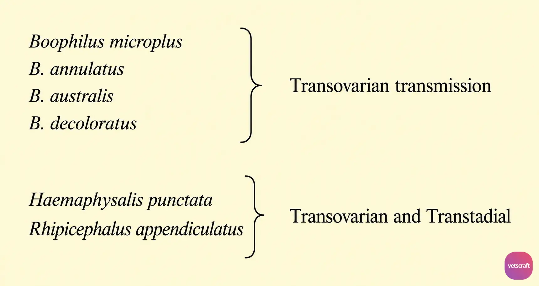
Transovarian transmission occurs in one-host ticks. Both transovarian and transtadial transmission occur in two- or three-host ticks.
Pathogenesis
Refer to the general pathogenesis section in the Babesia species post.
- Anemia
- Activation of kinins is more important.
- In Australia, this species is more pathogenic than B. bigemina, causing high mortality.
- Young animals show resistance to infection up to 1 year of age, whereas animals older than 2 years are more susceptible.
- Relapse (recurrence) may occur after 1½–2 years.
Clinical Signs
The incubation period is 4–10 days.
- High fever may persist for 7–10 days (104–106°F).
- Hemoglobinuria, anemia, diarrhea, icterus, and an increased heart rate.
- The cerebral form of babesiosis is very common in B. bovis. It is characterized by convulsions, incoordination, coma, followed by death.
- Brain capillaries are the predilection site for B. bovis.
Postmortem Lesions
- Congestion of the gray and white matter of the brain, with generalized dilation of capillaries containing parasitized RBCs.
- Perivascular, perineuronal, and interstitial edema throughout the brain and spinal cord.
Immunity
Premunition does not last for more than 2 years.
Diagnosis, treatment, and control are the same as those for B. bigemina.

