
TABLE OF CONTENTS
Canine Babesiosis in Dogs: Causes, Clinical Signs, Pathogenesis & Treatment
Canine babesiosis is a tick-borne hemoprotozoan disease of dogs caused by several species of the genus Babesia, primarily Babesia canis and Babesia gibsoni. The parasites invade erythrocytes (red blood cells), leading to hemolytic anemia, fever, jaundice, hemoglobinuria, and, in severe cases, multiple organ dysfunction or death. The disease is transmitted mainly by Rhipicephalus ticks, although blood transfusion, dog bites, and transplacental transmission have also been reported in certain cases.
This article provides a comprehensive overview of canine babesiosis, including the causative species, morphology, life cycle, pathogenesis, clinical signs, postmortem lesions, immunity, and treatment.
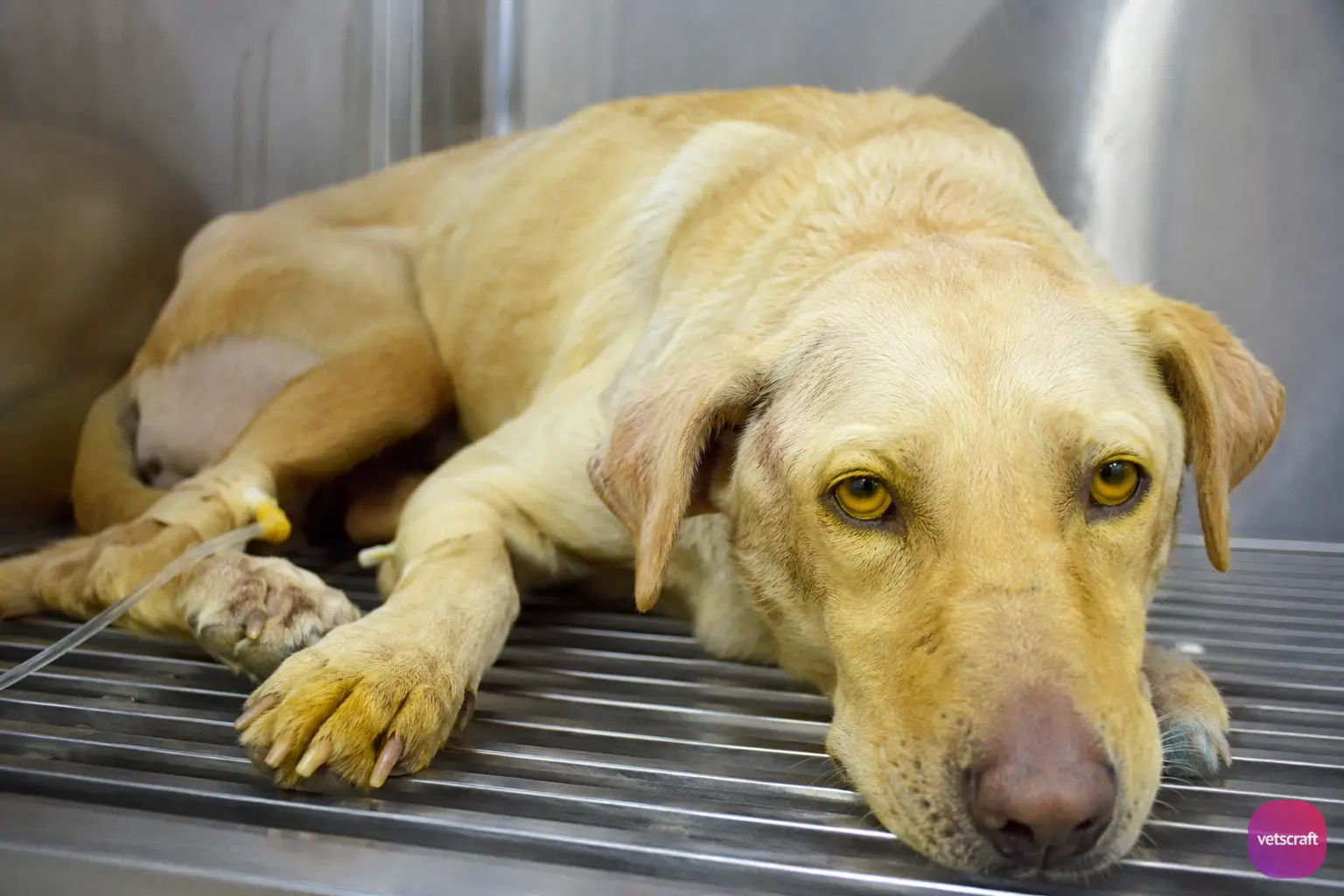
In canines (dogs), the following species of Babesia cause disease:
- Babesia canis
- Babesia gibsoni
- Babesia vogeli
Babesia canis
- Host: Dog, wolf, coyote, jackal, and fox
- Disease: Babesiosis in dogs is also known as biliary fever, malignant jaundice, canine babesiosis, and nambiuvu.
- Distribution: It is common in many tropical countries.
Morphology
- The merozoites are large, pyriform in shape, 4–5 µm long, and contain a vacuole. In the erythrocytes, they lie at an angle to one another.
- Pleomorphism may be seen in the merozoites, including amoeboid and ring forms.
- Multiple infections of erythrocytes are common; up to 16 merozoites may be found in a single RBC.
- Organisms may also be found in endothelial cells of the lungs and liver, and in macrophages due to erythrophagocytosis.
Life Cycle
Vectors: Rhipicephalus sanguineus. In this vector, Babesia is transmitted by both transovarial and transtadial transmission.
Pathogenesis
Although there is no age-specific susceptibility in canine babesiosis, young puppies are more susceptible. The incubation period is 10–21 days.
The clinical presentation of canine babesiosis is classified as follows:
Acute Disease
The first sign is fever, followed by anemia, icterus, loss of appetite, marked thirst, weakness, prostration, malaise, listlessness, reluctance to move, pale mucous membranes, jaundice, and death in many cases.
Hemoglobinuria may occur occasionally but is not usually present. There is enlargement of the spleen, the feces are markedly yellow in color, and a large amount of bilirubin is found in the urine.
Peracute Disease
There is hemoglobinuria, but the feces are not yellow; however, the animal frequently dies. The mode of death in canine babesiosis depends on the severity of the infection.
In severe cases, death usually occurs within 4–5 days due to acute respiratory failure, whereas in less severe cases, death may not occur rapidly, but the animals become progressively weaker and anemic, the extremities become cold, and death is associated with circulatory failure as a result of pulmonary edema.
Chronic Disease
This form is characterized by irregular fever, a capricious appetite, and marked loss of body condition.
Other Clinical Manifestations
Clinical signs such as catarrhal bronchitis, dyspnea, pneumonia, edema, ascites, stomatitis, and gastritis may be seen in dogs affected with babesiosis. Central nervous system involvement is common in B. canis.
In cerebral babesiosis, sudden death, incoordination, paresis, epileptic seizures, weakness of the limbs, and coma may occur.
Postmortem Lesions
- Enlargement of the liver and spleen
- The heart is pale and yellow.
- Kidneys show yellow nephrosis.
- Fat and mucous membranes are yellow.
Immunity
Premunity lasts for life.
Babesia gibsoni
- Host: Dog
- Vector: Rhipicephalus sanguineus
Morphology
- The merozoites are small and pleomorphic.
- They are usually oval or signet ring-shaped, resembling the piroplasms of Theileria annulata.
Life Cycle
The life cycle is similar to that of Babesia canis.
Pathogenesis and Clinical Signs
B. gibsoni causes a more chronic form of the disease. Affected dogs may have intermittent fever, anemia, and hemoglobinuria. In addition, epistaxis has been reported in dogs infected with B. gibsoni. Death may occur after several weeks or months.
Postmortem lesions include marked enlargement of the spleen and liver, but jaundice is not a frequent clinical sign.
Treatment
Berenil (diminazene aceturate) is effective in the treatment of canine babesiosis. Supportive therapy, including fluid therapy, blood transfusion (when indicated), and management of complications, may also be required in severely affected dogs.

