
Toxoplasma gondii: Life Cycle, Morphology, Pathogenesis, Mode of Infection & Classification
Toxoplasma gondii is an obligate intracellular protozoan parasite of worldwide importance that infects a wide range of warm-blooded animals, including humans. Members of the family Felidae serve as the definitive hosts, where the parasite undergoes sexual reproduction, while numerous mammals and birds act as intermediate hosts. T. gondii is the causative agent of toxoplasmosis, a zoonotic disease of significant veterinary and public health concern.
This article provides concise notes covering the parasite’s biological characteristics, developmental stages in definitive and intermediate hosts, transmission routes, and the pathogenesis of toxoplasmosis.
Parasite Overview
- Definitive Host: Members of the family Felidae, including the domestic cat, bobcat, leopard cat, jaguarundi, and ocelot.
- Intermediate Host: Warm-blooded animals, including humans, domestic animals, wild animals, and birds; over 200 species have been reported as intermediate hosts.
- Predilection Site: In the definitive host, several types of meronts and gamonts are found in the epithelial cells of the intestine, while tachyzoites and bradyzoites are found in the parenteral tissues. In the intermediate host, tachyzoites and bradyzoites (merozoites) are found in many types of cells, including neurons, microglia, endothelial cells, liver parenchymal cells, lungs, cardiac and skeletal muscles, fetal membranes, and leukocytes.
- Diseases Caused: Toxoplasmosis
- Distribution: Worldwide
Taxonomical Classification
- Kingdom: Protista
- Phylum: Apicomplexa
- Class: Conoidasida
- Subclass: Coccidiasina
- Order: Eucoccidiorida
- Family: Sarcocystidae
- Subfamily: Toxoplasmatinae
- Genus: Toxoplasma
- Species: Toxoplasma gondii
Morphology of Oocyst
- Oocysts are produced in the epithelial cells of the villi of the small intestine of cats and other felids.
- Unsporulated oocysts are spherical; after sporulation, they become subspherical (12.5 × 11 µm).
- No micropyle, residuum, or polar granule is present.
- The sporulated oocyst contains two sporocysts without a Stieda body. Each sporocyst contains four sporozoites.
- Sporulation Time: 2–3 days at 24°C.
Mode of Infection
Intermediate hosts (domestic animals, cattle, sheep, goats, wild animals, and birds) become infected by ingesting sporulated oocysts or infected meat containing tachyzoites or bradyzoites. In addition, congenital infection is common in intermediate hosts, particularly in humans.
Congenital toxoplasmosis in newborns occurs as a result of maternal infection during pregnancy. Infection can also be transmitted from one intermediate host to another through infected meat containing tachyzoites and bradyzoites.
Definitive hosts (felids) become infected by ingesting sporulated oocysts or infected meat containing tachyzoites or bradyzoites (pseudocysts) in the muscles and brain.
Life Cycle
Two types of life cycles exist in Toxoplasma gondii: the extraintestinal life cycle, which occurs in both definitive and intermediate hosts, and the intestinal life cycle, also called the enteroepithelial life cycle, which occurs only in the definitive host.
Extraintestinal Life Cycle
When sporulated oocysts are ingested by intermediate hosts or the definitive host (cat), sporozoites are released in the intestine.
These liberated sporozoites penetrate the intestinal wall. From there, they are carried by the blood or lymph to various parts of the body (parenteral tissues). Upon reaching the parenteral tissues, they invade various tissue types and multiply rapidly by endodyogeny.
These rapidly dividing merozoites are called tachyzoites. Tachyzoites are crescent-shaped with a centrally placed nucleus and an apical complex at the anterior end. Tachyzoite development usually occurs during acute infection and may be detected in the blood in severe cases.
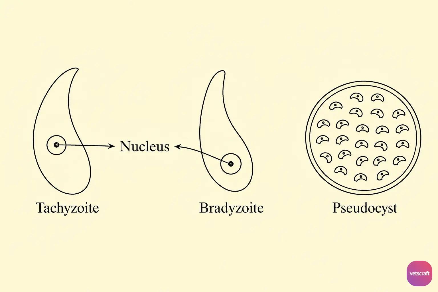
Tachyzoites, usually numbering approximately 8–16, accumulate within host cells and are referred to as groups, terminal colonies, or colony aggregate stages. Tachyzoites are less resistant to gastric secretions; therefore, they are considered a less important source of infection.
As the infection becomes chronic, tachyzoites enter other cells, such as those in the brain, heart, and skeletal muscles, where they multiply very slowly. These slowly multiplying merozoites are called bradyzoites.
Bradyzoites accumulate in large numbers within a host cell and form a cyst by stimulating the host cell to produce a tough surrounding wall, called a pseudocyst or zoitocyst.
Pseudocyst formation occurs in the brain and usually coincides with the development of immunity against new infection.
If immunity wanes, bradyzoites are released, which can boost immunity to its previous level (premunition). Cell-mediated immunity plays an important role in toxoplasmosis. Pseudocyst formation marks the end of the life cycle in the intermediate host.
Intestinal Life Cycle (Enteroepithelial Life Cycle)
This type of life cycle occurs only in definitive hosts (cats and other felids). When meat containing pseudocysts is ingested, bradyzoites are released into the intestine.
The released bradyzoites enter the intestinal epithelial cells and produce five developmental stages: A, B, C, D, and E.
- A: Multiplication by endodyogeny
- B: Endodyogeny + Endopolygeny
- C: Schizogony
- D: Endodyogeny + Schizogony
- E: Schizogony
Type E differentiates into macrogamonts and microgamonts in the intestinal epithelial cells. The microgamonts produce 12–32 triflagellated microgametes, which fuse with the macrogamete, resulting in the formation of a zygote.
An oocyst wall forms around the zygote, and it is excreted in the feces as an unsporulated oocyst.
The prepatent period in cats depends on the stage ingested:
- Bradyzoites (cyst): 2–7 days
- Tachyzoites: 7–10 days
- Oocysts: 1–2 weeks
Pathogenesis
Sexual stages are not pathogenic in the definitive host. Asexual stages found in the intermediate host may cause disease ranging from inapparent infection to acute fatal disease. Therefore, asymptomatic toxoplasmosis is common.
In humans, congenital toxoplasmosis is common in infants. It is characterized by encephalitis, rash, jaundice, hepatomegaly, chorioretinitis, hydrocephalus, and microcephaly, with a high mortality rate.
Acquired toxoplasmosis is manifested in four different forms:
- Type I is characterized by lymphadenopathy and fever. The onset of clinical signs may be acute, with chills and fever lasting 2–4 weeks. Enlargement of the lymph nodes, sore throat, malaise, and fatigue may persist even after recovery.
- Type II is a typhus-like exanthematous disease characterized by atypical pneumonia, myocarditis, and meningoencephalitis. Lymphadenopathy may or may not be present, but the condition is usually fatal.
- Type III is the cerebrospinal form, characterized by fever, encephalitis, convulsions, delirium, lymphadenopathy, mononuclear pleocytosis, and eventual death.
- Type IV is the ophthalmic form, which is the most common form of acquired toxoplasmosis, characterized by chronic chorioretinitis.

