
Toxoplasmosis in Animals: Pathogenesis, Diagnosis, Treatment & Control
Toxoplasmosis in animals is a globally distributed protozoal disease caused by Toxoplasma gondii. It affects a wide range of domestic and wild animals, with clinical manifestations varying depending on the host species, age, immune status, and stage of infection. While many infections remain subclinical, toxoplasmosis can cause significant reproductive losses, neurological disorders, and systemic illness in susceptible animals.
This article provides a concise overview of the pathogenesis of toxoplasmosis, laboratory diagnosis, the Sabin-Feldman dye test, available treatment options, preventive measures, and the use of the Toxovac vaccine in sheep.
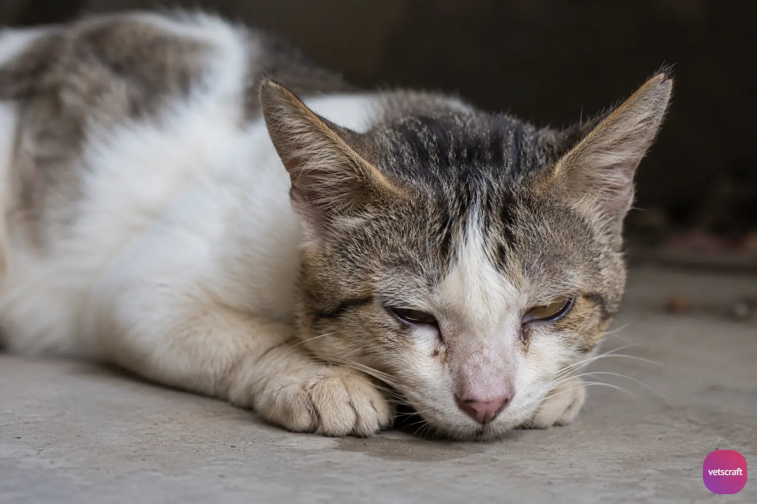
Pathogenesis of Toxoplasmosis
Pathogenesis in domestic animals is similar to that in humans. However, clinical signs may vary from species to species.
- Cat: There is enteritis with ulcers, enlargement of the mesenteric lymph nodes, CNS degeneration, and diarrhea.
- Cattle: Affected animals have dyspnea, nasal discharge, sneezing, and coughing. There is trembling and shaking of the head.
- Sheep: Toxoplasmosis in sheep is an economically important disease because it causes abortion. Abortion occurs only if the ewe becomes infected during the early stage of pregnancy. As toxoplasmosis causes a high rate of abortions, it is referred to as abortion storms in sheep.
- Pig: There will be fever, shivering, incoordination, relaxation of the abdominal muscles, and diarrhea.
Diagnosis of Toxoplasmosis
Diagnosis based on clinical signs is very difficult because the disease is mostly asymptomatic.
- HAT
- ELISA
- Indian ink immunoreaction
- MAT (Modified Agglutination Test)
- Latex agglutination
- IFA
The most reliable method of diagnosis is isolation of the parasite by inoculating the suspected material into mice through the intraperitoneal route. If the material is positive, the mice will develop ascites, and tachyzoites can be demonstrated in smears prepared from the peritoneal fluid and cut surfaces of the lungs, liver, and spleen, while bradyzoites can be seen in brain impression smears.
Sabin-Feldman Dye Test
- Principle: Toxoplasma antibodies and the accessory factor complement properdin modify (kill) the living tachyzoites and bradyzoites of Toxoplasma. As a result, they fail to stain with methylene blue at pH 11.
- Disadvantage: An experienced technician is necessary for the interpretation of the dye test.
The cytoplasm and nucleus of the tachyzoites and bradyzoites of T. gondii stain deeply with methylene blue after incubation with normal serum (negative), whereas if they have been incubated with serum containing Toxoplasma antibodies, they do not take up the stain except in the nuclear endosome.
Positivity can be determined by the highest serum dilution that is able to modify 50% of the tachyzoites and bradyzoites of Toxoplasma.
Treatment
- No satisfactory treatment is available for toxoplasmosis.
- Pyrimethamine 1 mg/kg is effective.
- Pyrimethamine 1 mg/kg + Sulphadiazine 120 mg/kg.
- Clindamycin 13.5 mg/kg three times daily for 10 days.
- Toltrazuril is effective for toxoplasmosis in dogs and cats.
Control
- Avoid contact with stray cats and wild animals that have been fed an uncontrolled diet.
- Avoid eating raw meat.
- Provide well-cooked meat to cats.
- Pregnant women should not handle or care for cats.
- Avoid contact with contaminated soil.
- Use disposable gloves while handling cats.
- Vaccination of sheep.
Toxovac
Toxovac is a vaccine developed against toxoplasmosis in sheep. It contains living tachyzoites of the S–48 strain of T. gondii. Young sheep can be vaccinated from 5 months of age onward, and older animals should be vaccinated 4 months prior to mating. The dose is 2 mL of diluted vaccine, administered intramuscularly.
Warning
- Pregnant animals should not be vaccinated.
- Pregnant women should not handle the vaccine.

