
Sarcocystis cruzi: Morphology, Life Cycle, Pathogenesis, Diagnosis & Treatment
Sarcocystis cruzi is an obligate intracellular protozoan parasite of the family Sarcocystidae that follows a two-host (predator–prey) life cycle. Dogs and other canids serve as the definitive hosts, while cattle are the intermediate hosts. Although infection in definitive hosts is usually subclinical, S. cruzi can cause significant disease in cattle, resulting in economic losses due to reduced productivity, abortion, and mortality.
This article provides a comprehensive overview of Sarcocystis cruzi, including its morphology, taxonomical classification, transmission, life cycle, pathogenesis, diagnosis, treatment, and control.
Parasite Overview
- Definitive Host: Dogs, wolves, coyotes, foxes, and hyenas
- Intermediate Host: Cattle
- Predilection Site: Sarcocysts in the muscles of the intermediate host. Sexual stages and oocysts in the intestine of the definitive host.
- Disease Caused: Sarcocystosis and Sarcosporidiosis
- Distribution: Worldwide
Taxonomical Classification
- Kingdom: Animalia
- Phylum: Apicomplexa
- Class: Conoidasida
- Subclass: Coccidiasina
- Order: Eucoccidiorida
- Family: Sarcocystidae
- Genus: Sarcocystis
- Species: Sarcocystis cruzi
Morphology of Oocyst
Oocysts resemble those of Isospora, contain 2 sporocysts, each containing 4 sporozoites. Oocysts are dumbbell-shaped and measure 19–21 × 15–18 µm, with no micropyle, polar granule, or Stieda body.
Sporocysts are ellipsoidal, with one side flatter than the other. The oocyst wall is smooth, colorless, and pale yellow. Sporozoites are banana-shaped, with one end rounded and the other pointed.
Transmission
The intermediate host becomes infected by ingesting sporulated oocysts while grazing, and definitive hosts become infected by ingesting muscle cysts containing bradyzoites.
Meronts and metrocytes are not infective to the definitive host, but tachyzoites are able to infect other intermediate hosts.
Life Cycle
When sporocysts or oocysts are ingested by the intermediate host, sporozoites are released in the intestine. The liberated sporozoites penetrate the intestinal epithelium and reach the bloodstream.
They are then carried by the bloodstream to various parts of the body, where they invade the endothelial cells of small blood vessels and undergo multiplication by endodyogeny.
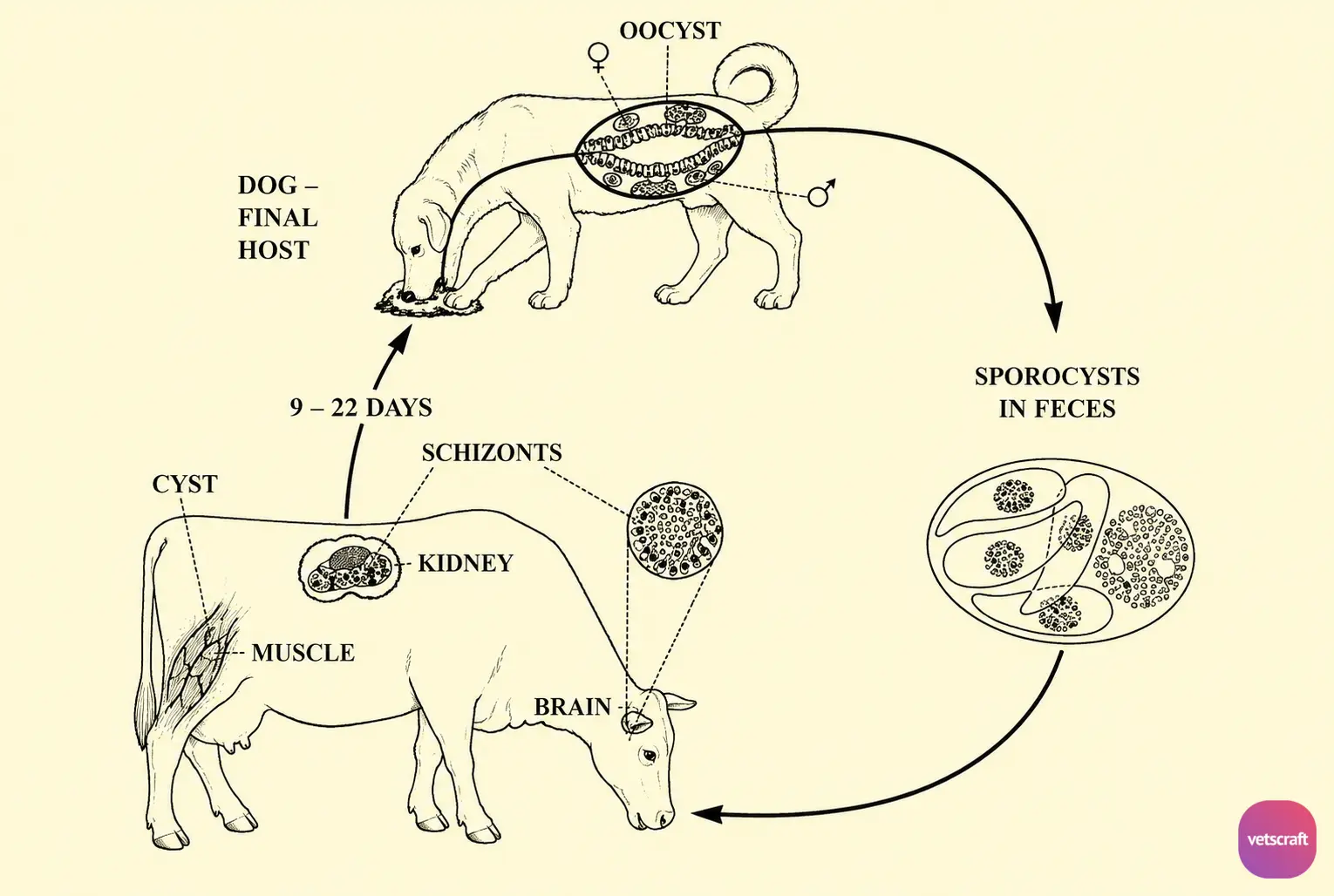
The first- and second-generation meronts occur in the endothelial cells and contain approximately 200 banana-shaped tachyzoites. In acute infections, tachyzoites may be found in the blood.
The third-generation merogony occurs in circulating lymphocytes, and the tachyzoites of the third-generation meronts enter the striated muscles, cardiac muscle, and brain, where they produce the final-generation meront called a sarcocyst or Miescher’s tubule within 45 days of infection. These appear as boiled rice grains in the muscles.
Structure of Sarcocysts
Sarcocysts are found in the striated muscles, cardiac muscle, and rarely in the brain, but are more commonly found in the esophagus and tongue. Sarcocysts are 1 cm long and 1–2 mm in diameter, elongated, cylindrical, or spindle-shaped, and lie between the muscle fibers. They have a definite wall composed of a primary layer and a secondary layer.
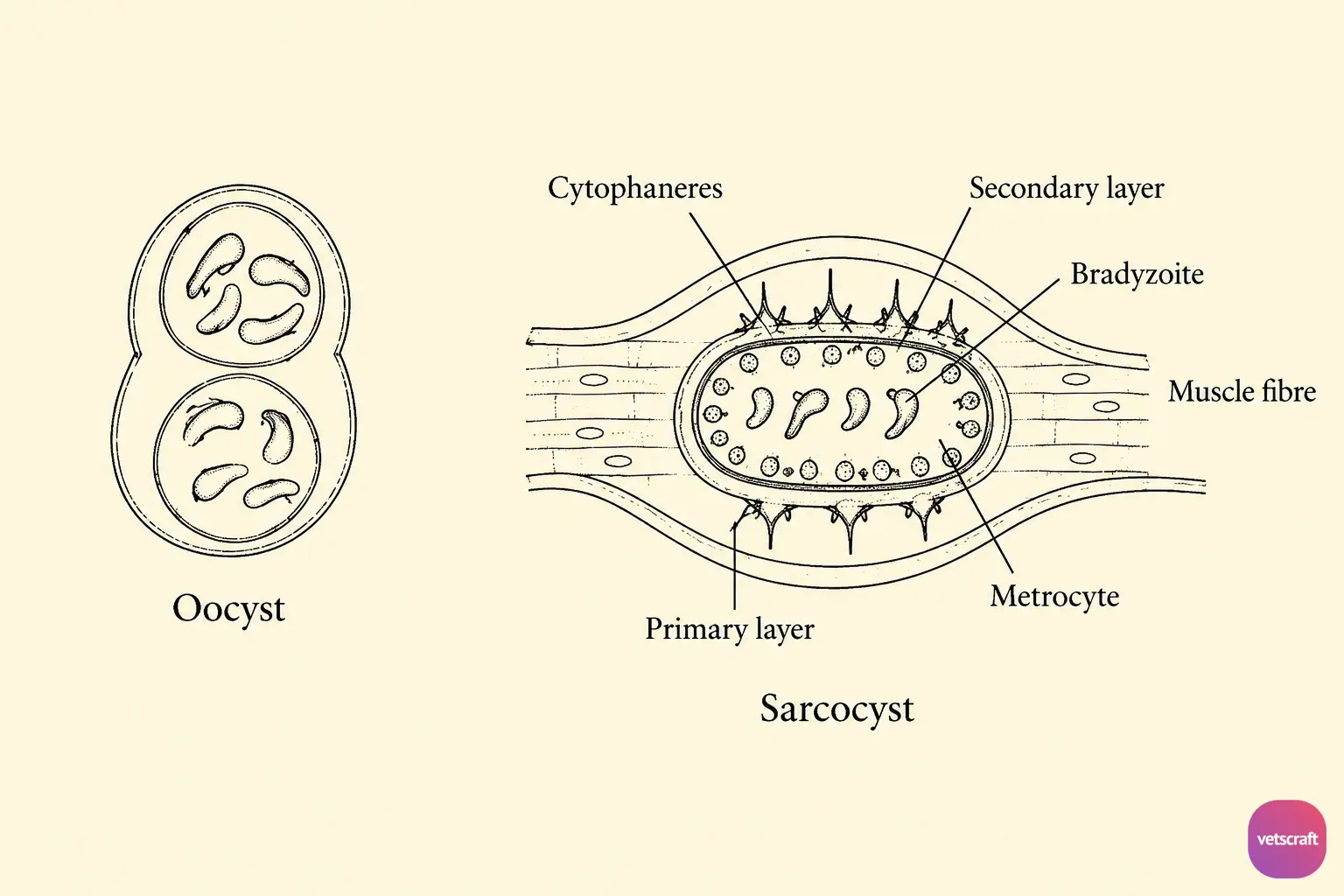
The primary layer bears numerous radial spines called cytophaneres, which radiate into the muscle fibers. The secondary layer has trabeculae or septa that divide the sarcocyst into numerous compartments.
There are two distinct regions in the sarcocyst. The peripheral region is occupied by globular metrocytes (promerozoites), which later undergo repeated division by endodyogeny to form intermediate stages and then bradyzoites (Rainey’s corpuscles) or zoitocysts. The bradyzoites are bean- or sickle-shaped and resemble coccidian merozoites. Only bradyzoites are infective to the definitive host.
Infection of the definitive host occurs when infected meat containing zoitocysts or bradyzoites is ingested. In the stomach, the cyst wall is digested, releasing the bradyzoites. These liberated bradyzoites then penetrate the lamina propria of the small intestine, where they differentiate into macrogamonts and microgamonts, which in turn produce gametes.
The male gametes penetrate the female gametes, resulting in zygote formation, which then becomes an oocyst. The oocysts of Sarcocystis sporulate in the lamina propria of the intestine. Since the oocyst wall is thin, it may rupture while passing through the intestine; thus, sporocysts rather than oocysts are normally passed in the feces.
Pathogenesis
Sarcocystis cruzi does not produce significant pathogenesis in the definitive host, but it causes severe disease in cattle. The pathogenesis is mainly due to tachyzoites and meronts in the endothelial cells, whereas intact sarcocysts and bradyzoites do not produce any pathogenic effects.
The sarcocyst contains a powerful endotoxin known as sarcocystin, which is highly toxic and may cause damage to the CNS, heart, adrenal glands, liver, intestinal wall, etc. Clinical signs of the disease include mild fever, diarrhea, and, in severe cases, death.
In cattle, the disease is characterized by anorexia, cachexia, weight loss, anemia, increased heart rate, reduced milk yield, dyspnea, and loss of hair at the tip of the tail, followed by abortion and death. This condition is called Dalmeny disease. Chronic infection is characterized by emaciation, submandibular edema, and exophthalmia.
Postmortem Lesions
Ecchymoses occur in various organs.
Immunity
- There is no immunity in the definitive host.
- In the intermediate host, some degree of immunity may develop.
Diagnosis
In the Definitive Host
Examination of feces for the presence of oocysts or sporocysts. Intestinal scrapings can be examined for gamonts.
In the Intermediate Host
Sarcocystis can be diagnosed only during postmortem examination of dead animals and routine meat inspection of carcasses in slaughterhouses. Sarcocysts are found in the striated muscles, tongue, esophagus, and heart, where they appear as boiled rice grains.
- Histopathological sections of muscles show various developmental stages, viz., tachyzoites, bradyzoites, and metrocytes.
- IHA
Treatment
- Dogs do not require any treatment, but treatment protocols used for Isospora spp. can be followed.
- For Cattle: Amprolium at 100 mg/kg body weight.
Control
- Proper disposal of dog feces.
- Prevent contamination of cattle feed and water/grazing areas with dog feces.
- Provide well-cooked meat to dogs.
- Proper disposal of carcasses.
- Freeze meat to kill the bradyzoites.

