
Hepatozoon canis in Animals: Life Cycle, Transmission, Pathogenesis, Diagnosis & Treatment
Hepatozoon canis is a tick-borne protozoan parasite that primarily infects dogs, although it has also been reported in cats, jackals, and hyenas. Unlike most tick-borne pathogens, transmission occurs when a susceptible animal ingests an infected tick rather than through a tick bite. After ingestion, the parasite undergoes complex developmental stages in both the vertebrate host and the tick vector. Although many infected animals remain asymptomatic, severe infections can cause fever, anemia, weight loss, splenomegaly, and other systemic signs.
This article provides an overview of the taxonomy, morphology, life cycle, transmission, pathogenesis, diagnosis, and treatment of Hepatozoon canis.
Parasite Overview
- Protozoan Parasite: Hepatozoon canis
- Hosts: Dogs, cats, jackals, and hyenas
- Predilection Site:
- Schizogony occurs in the endothelial cells of the liver, spleen, and bone marrow, while gametogony occurs in the neutrophils of dogs.
- Sporogony occurs in the gut epithelial cells of ticks, and oocysts develop in the haemocoel cavity.
- Vector: Brown dog tick: Rhipicephalus sanguineus (transmitted by nymphs and adults)
- Distribution: East, Central, and North Africa; common in India.
Taxonomical Classification
- Kingdom: Protista
- Phylum: Apicomplexa
- Class: Sporozoea
- Order: Eucoccidiorida
- Suborder: Adeleidea
- Family: Haemogregarinidae
- Genus: Hepatozoon
- Species: Hepatozoon canis
Structure of Gamonts
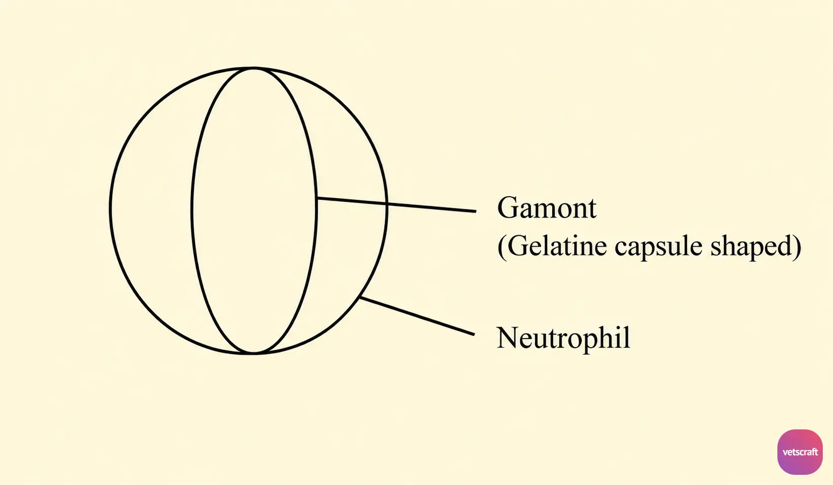
Gamonts are found in neutrophils. They are elongate, rectangular bodies (8–12 × 3–6 µm), surrounded by a delicate capsule (gelatin capsule), which stains pale blue with a dark reddish nucleus and contains a number of pink granules in the cytoplasm.
Transmission and Life Cycle
Development in the Definitive Host
Dogs become infected by ingesting infected ticks. The ingested ticks are digested, leading to the release of sporozoites in the intestine. The liberated sporozoites penetrate the intestinal wall, from where they are carried via the bloodstream to different tissues such as the liver, spleen, bone marrow, and other tissue cells.
The sporozoites then enter tissue cells, where they multiply to produce two types of meronts, namely macromeronts and micromeronts.
The macromeronts produce a small number of large merozoites, whereas the micromeronts produce a large number of small merozoites. After several generations of asexual reproduction, the small merozoites enter the neutrophils and differentiate into macrogamonts and microgamonts.
Further development of the parasite occurs in the tick vector following a blood meal from infected dogs.
Development in the Tick Vector
When ticks feed on infected dogs, gamonts are ingested along with the blood meal. These gamonts leave the neutrophils in the lumen of the tick gut, after which the macro- and microgamonts become attached in pairs, a process known as syzygy.
Each microgamont produces two non-flagellated microgametes, one of which fertilizes the macrogamete to produce a zygote. This motile zygote is called an ookinete. The ookinetes then penetrate the intestinal wall and reach the haemocoel cavity, where they develop into oocysts. As the oocysts mature, sporoblasts and sporocysts are formed, each of which produces 16 sporozoites.
Pathogenesis
H. canis is not highly pathogenic in dogs. It may be found in apparently healthy dogs without any clinical signs; however, severe infections may cause irregular fever, anemia, emaciation, splenomegaly, and lumbar paralysis.
Diagnosis
Examination of blood smears for the presence of gamonts in neutrophils.
Treatment
Tetracycline may be administered.

