TABLE OF CONTENTS
Fasciola (Liver Fluke): Morphology, Life Cycle, Pathogenesis, Diagnosis and Control
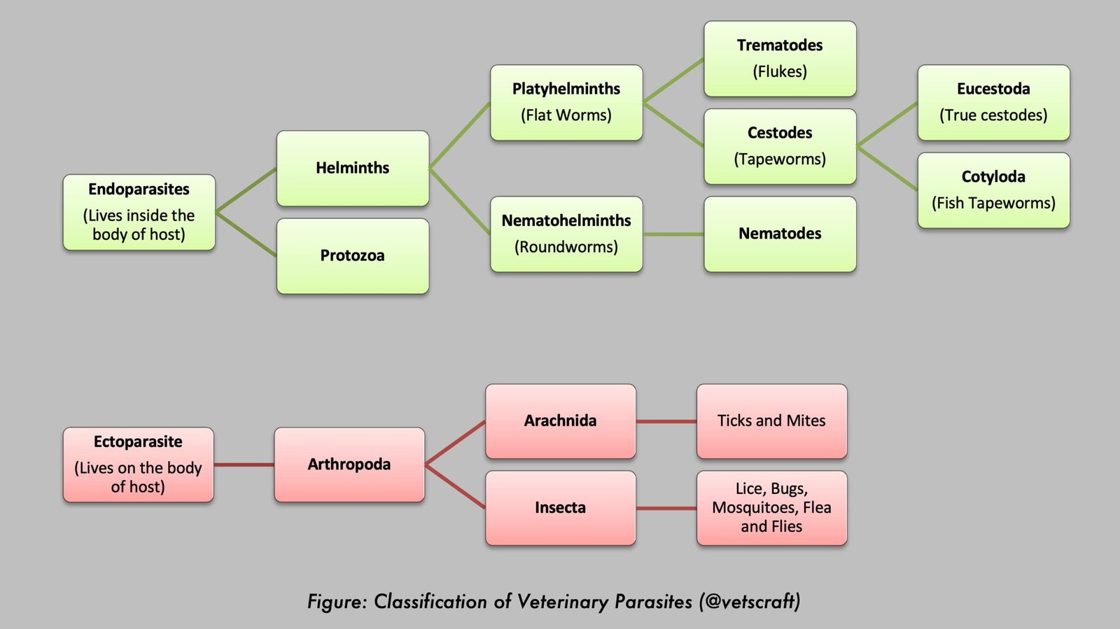
Fasciola spp. are parasitic trematodes commonly known as liver flukes. The two major species of veterinary importance are Fasciola hepatica and Fasciola gigantica. These parasites infect a wide range of domestic and wild mammals and cause fasciolosis, a disease associated with liver damage, reduced productivity, weight loss, and economic losses in livestock production.
| Feature | Details |
|---|---|
| Common Name | Liver Fluke |
| Scientific Name | Fasciola spp. |
| Major Species | Fasciola hepatica and Fasciola gigantica |
| Disease Caused | Fasciolosis (Liver Fluke Disease) |
| Family | Fasciolidae |
| Intermediate Host | Aquatic snails (Lymnaea spp.) |
| Definitive Hosts | Cattle, buffalo, sheep, goats, and other mammals, including humans |
| Predilection Site | Liver parenchyma and bile ducts |
| Infective Stage | Metacercaria |
| Diagnostic Stage | Eggs in feces |
| Mode of Transmission | Ingestion of metacercariae on contaminated vegetation or water |
| Distribution | Worldwide |
Taxonomical Classification of Fasciola
- Family: Fasciolidae
- Genus: Fasciola
- Species: Fasciola gigantica, Fasciola hepatica
- Common Name: Liver fluke
- Disease: Fasciolosis, liver rot, fluke cirrhosis, clay-pipe cirrhosis, pipe-stem liver, and gallstone formation.
- Distribution: Worldwide
- Hosts: Cattle, buffalo, sheep, goats, and humans.
- Location: Bile ducts and liver parenchyma
- Intermediate Hosts (I/H): Aquatic snails – Lymnaea acuminata, L. rufescens, L. auricularia, and L. natalensis
Morphology of F. gigantica
- Leaf-shaped and large in size.
- Measures 25–75 mm in length and about 12 mm in breadth; broader anteriorly than posteriorly.
- The anterior cone is small.
- Shoulders are indistinct (not prominent).
- The cuticle bears spines (spinose tegument).
- The body is relatively transparent.
- Oral sucker (OS) and ventral sucker (VS) are present. The VS is situated at the level of the shoulder and is larger than the OS.
- The intestinal caeca are branched and diverticulated both externally and internally.
- Two follicular, branched testes are located in the middle region.
- A single branched ovary with a coiled uterus lies anterior to the testes.
- The genital opening is located anterior to the ventral sucker.
- Numerous vitellaria are present laterally, along with two transverse vitelline ducts.
Morphology of F. hepatica
- Comparatively smaller (approximately 30 × 13 mm) than F. gigantica.
- Distinct anterior cone present.
- Prominent shoulders.
- OS and VS are of similar size.
- Intestinal caeca are branched and diverticulated only externally.
- Testes are located in the posterior region.
- The most common liver fluke in temperate regions.
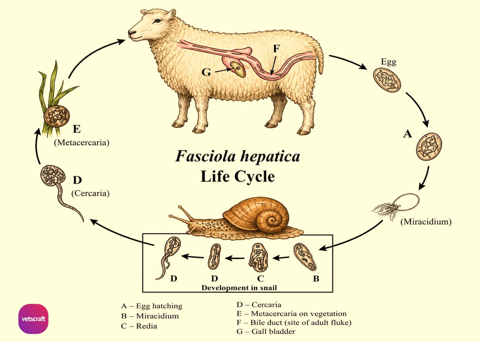
Life Cycle
Egg
The egg of Fasciola is of the composite type. It is large, measuring approximately 156–197 × 90–104 µm, oval in shape, and yellowish in color with an indistinct operculum. It contains yolk cells and hexagonal-shaped germ cells.
Eggs are passed in the feces of the host. They can survive for 2–3 months in moist feces but are rapidly destroyed by desiccation within a few hours to days. Miracidia develop only after the eggs have been laid.
Development in the Invertebrate Host (Snail I/H)
Eggs hatch in approximately 10–12 days at 26°C, depending on temperature, and release miracidia.
Miracidium
The miracidium is broad anteriorly, possesses a small papiliform protrusion, a ciliated tegument, and a pair of eyespots. It must reach the snail host within approximately 3 hours of hatching.
The miracidium actively penetrates an aquatic snail (e.g., L. acuminata), sheds its ciliated covering, and develops into a mother sporocyst. Each sporocyst gives rise to 1–6 rediae through polyembryony.
Redia
The redia possesses a circular thickening behind the pharynx and a pair of blunt processes near the beginning of the posterior quarter of the body. It measures approximately 1–3 mm in length. Usually, rediae produce cercariae, although daughter rediae may develop under unfavorable conditions.
Cercaria
The cercaria is of the Gymnocephalus type. It lacks spines and eyespots but possesses dark granular cystogenous glands in the lateral part of the body and a long tail. Development within the snail takes approximately 5–7 weeks under favorable conditions.
Within a few minutes to 2 hours after leaving the snail, the cercaria settles on grass blades or other herbage just below the water level (submerged vegetation) and sheds its tail.
It secretes a cyst wall through the cystogenous glands and forms a spherical cyst known as a metacercaria. A small number may encyst on the water surface and subsequently sink to the bottom.
The metacercaria is the infective stage for the definitive host (D/H). It appears pearly or milky white and is approximately the size of a pinhead. Infection of a snail by a single miracidium may produce more than 600 metacercariae. Under laboratory conditions, metacercariae can survive for up to one year.
At 70% relative humidity, metacercariae can survive for 270–340 days on herbage, approximately 8 months on moist hay, and 35–57 days in silage. Under normal conditions, they survive for 3–6 months but die rapidly during hot, dry, and sunny weather.
Development in the Vertebrate Host (Definitive Host)
The definitive host acquires infection by ingesting metacercariae on contaminated herbage or in drinking water. Following ingestion, excystation occurs in the duodenum. Within 24 hours of infection, the majority of immature flukes are found in the abdominal and peritoneal cavities.
By 4–6 days post-infection, most immature flukes penetrate the liver capsule and begin migrating through the liver parenchyma.
Although young flukes may occasionally reach the liver through the bloodstream, this route is uncommon. The usual route is through the peritoneal cavity, and migration within the liver may continue for up to 5–6 weeks.
From the 7th week onward, the flukes enter the bile ducts and attain sexual maturity. Some flukes may also be found in the gallbladder.
During the 8th week of infection, eggs begin to appear in the feces.
Occasionally in cattle, immature flukes may be found in other organs such as the lungs. In pregnant animals, the parasite may also occur in the fetus.
The minimum period required to complete one life cycle (egg to egg) is approximately 17–18 weeks in F. gigantica under favorable conditions.
The longevity of Fasciola in untreated sheep may extend for several years, whereas in cattle it is generally less than one year.
Pathogenesis
Pathogenesis depends on the number of metacercariae ingested and the immune status of the host. Little to no damage occurs in the intestinal wall or peritoneal cavity during migration.
Lesions occur primarily in the liver. Pathogenesis is mainly associated with immature flukes migrating through the liver parenchyma and adult flukes residing in the bile ducts.
Ovine and Bovine Fasciolosis
Fasciolosis affects both sheep (ovine fasciolosis) and cattle (bovine fasciolosis), although the disease manifestations differ between species. Sheep are generally more susceptible and may develop acute, subacute, or chronic disease due to extensive migration of immature flukes through the liver parenchyma. In cattle, fasciolosis is usually chronic, characterized by progressive weight loss, anemia, reduced milk production, and decreased productivity.